 Movie
Movie Controller

[English] 日本語
 Yorodumi
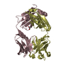
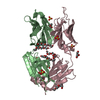
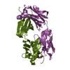
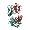
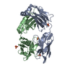
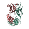
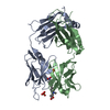
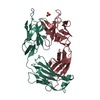
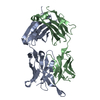
- PDB-6wrp: Crystal Structure of PI3-E12 Fab, An Antibody Against Human Parai... -
+ Open data

- Basic information
| Entry | Database: PDB / ID: 6wrp | ||||||
|---|---|---|---|---|---|---|---|
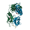
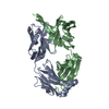
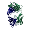
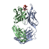
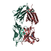
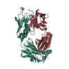
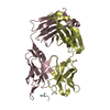
| Title | Crystal Structure of PI3-E12 Fab, An Antibody Against Human Parainfluenza Virus Type III | ||||||
 Components Components |
| ||||||
 Keywords Keywords | IMMUNE SYSTEM / antibody / FAB | ||||||
| Function / homology | Immunoglobulins / Immunoglobulin-like / Sandwich / Mainly Beta / AMMONIUM ION / DI(HYDROXYETHYL)ETHER Function and homology information Function and homology information | ||||||
| Biological species |  Homo sapiens (human) Homo sapiens (human) | ||||||
| Method |  X-RAY DIFFRACTION / MOLECULAR REPLACEMENT / Resolution: 2.08 Å X-RAY DIFFRACTION / MOLECULAR REPLACEMENT / Resolution: 2.08 Å | ||||||
 Authors Authors | Weidle, C. / Pancera, M. | ||||||
| Funding support |  United States, 1items United States, 1items
| ||||||
 Citation Citation | Journal: Mabs / Year: 2021 Title: Protective antibodies against human parainfluenza virus type 3 infection. Authors: Boonyaratanakornkit, J. / Singh, S. / Weidle, C. / Rodarte, J. / Bakthavatsalam, R. / Perkins, J. / Stewart-Jones, G.B.E. / Kwong, P.D. / McGuire, A.T. / Pancera, M. / Taylor, J.J. | ||||||
| History |
|
- Structure visualization
| Structure viewer | Molecule: MolmilJmol/JSmol |
|---|
- Downloads & links
-Download
| PDBx/mmCIF format | 6wrp.cif.gz | 192.3 KB | Display | PDBx/mmCIF format |
|---|---|---|---|---|
| PDB format | pdb6wrp.ent.gz | 141.1 KB | Display | PDB format |
| PDBx/mmJSON format | 6wrp.json.gz | Tree view | PDBx/mmJSON format | |
| Others |  Other downloads Other downloads |
-Validation report
| Arichive directory | https://data.pdbj.org/pub/pdb/validation_reports/wr/6wrpftp://data.pdbj.org/pub/pdb/validation_reports/wr/6wrp | HTTPS FTP |
|---|
-Related structure data
| Related structure data | 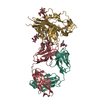 6mftS S: Starting model for refinement |
|---|---|
| Similar structure data |


-Links
 PDBj
PDBj


- Assembly
| Deposited unit | 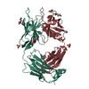
| ||||||||||||
|---|---|---|---|---|---|---|---|---|---|---|---|---|---|
| 1 |
| ||||||||||||
| Unit cell |
|
-Components
-Antibody , 2 types, 2 molecules HL
| #1: Antibody | Mass: 24763.932 Da / Num. of mol.: 1 Source method: isolated from a genetically manipulated source Source: (gene. exp.) Homo sapiens (human) / Cell (production host): 293 / Production host: Homo sapiens (human) |
|---|---|
| #2: Antibody | Mass: 23772.463 Da / Num. of mol.: 1 Source method: isolated from a genetically manipulated source Source: (gene. exp.) Homo sapiens (human) / Production host: Homo sapiens (human) |
-Non-polymers , 5 types, 350 molecules 


| #3: Chemical | ChemComp-PEG /  Mass: 106.120 Da / Num. of mol.: 4 / Source method: obtained synthetically / Formula: C4H10O3 Mass: 106.120 Da / Num. of mol.: 4 / Source method: obtained synthetically / Formula: C4H10O3#4: Chemical |  Mass: 62.068 Da / Num. of mol.: 2 / Source method: obtained synthetically / Formula: C2H6O2 Mass: 62.068 Da / Num. of mol.: 2 / Source method: obtained synthetically / Formula: C2H6O2#5: Chemical | ChemComp-SO4 /  Mass: 96.063 Da / Num. of mol.: 4 / Source method: obtained synthetically / Formula: SO4 Mass: 96.063 Da / Num. of mol.: 4 / Source method: obtained synthetically / Formula: SO4#6: Chemical |  Mass: 18.038 Da / Num. of mol.: 2 / Source method: obtained synthetically / Formula: H4N Mass: 18.038 Da / Num. of mol.: 2 / Source method: obtained synthetically / Formula: H4N#7: Water | ChemComp-HOH / | Mass: 18.015 Da / Num. of mol.: 338 / Source method: isolated from a natural source / Formula: H2O |
|---|
-Details
| Has ligand of interest | N |
|---|---|
| Has protein modification | Y |
-Experimental details
-Experiment
| Experiment | Method: X-RAY DIFFRACTION / Number of used crystals: 1 |
|---|
- Sample preparation
| Crystal | Density Matthews: 2.09 Å3/Da / Density % sol: 41.27 % |
|---|---|
| Crystal grow | Temperature: 295 K / Method: vapor diffusion, hanging drop Details: 2.2M Ammonium Sulfate, 2.2% PEG 400, 0.11M Hepes pH 7.5 |
-Data collection
| Diffraction | Mean temperature: 100 K / Serial crystal experiment: N |
|---|---|
| Diffraction source | Source: ROTATING ANODE / Type: RIGAKU MICROMAX-007 HF / Wavelength: 1.5418 Å |
| Detector | Type: RIGAKU SATURN 944+ / Detector: CCD / Date: Feb 16, 2019 |
| Radiation | Monochromator: M / Protocol: SINGLE WAVELENGTH / Monochromatic (M) / Laue (L): M / Scattering type: x-ray |
| Radiation wavelength | Wavelength: 1.5418 Å / Relative weight: 1 |
| Reflection | Resolution: 2.08→50 Å / Num. obs: 24469 / % possible obs: 98 % / Redundancy: 6.5 % / Biso Wilson estimate: 26.41 Å2 / CC1/2: 0.997 / Net I/σ(I): 16.01 |
| Reflection shell | Resolution: 2.09→2.13 Å / Num. unique obs: 2026 / CC1/2: 0.905 |
- Processing
| Software |
| |||||||||||||||||||||||||||||||||||||||||||||||||||||||||||||||
|---|---|---|---|---|---|---|---|---|---|---|---|---|---|---|---|---|---|---|---|---|---|---|---|---|---|---|---|---|---|---|---|---|---|---|---|---|---|---|---|---|---|---|---|---|---|---|---|---|---|---|---|---|---|---|---|---|---|---|---|---|---|---|---|---|
| Refinement | Method to determine structure: MOLECULAR REPLACEMENT Starting model: 6MFT Resolution: 2.08→23.79 Å / SU ML: 0.2547 / Cross valid method: FREE R-VALUE / σ(F): 1.33 / Phase error: 26.3616 Stereochemistry target values: GeoStd + Monomer Library + CDL v1.2
| |||||||||||||||||||||||||||||||||||||||||||||||||||||||||||||||
| Solvent computation | Shrinkage radii: 0.9 Å / VDW probe radii: 1.11 Å / Solvent model: FLAT BULK SOLVENT MODEL | |||||||||||||||||||||||||||||||||||||||||||||||||||||||||||||||
| Displacement parameters | Biso mean: 30.24 Å2 | |||||||||||||||||||||||||||||||||||||||||||||||||||||||||||||||
| Refinement step | Cycle: LAST / Resolution: 2.08→23.79 Å
| |||||||||||||||||||||||||||||||||||||||||||||||||||||||||||||||
| Refine LS restraints |
| |||||||||||||||||||||||||||||||||||||||||||||||||||||||||||||||
| LS refinement shell |
|
