 Movie
Movie Controller

[English] 日本語
 Yorodumi
- PDB-2v7n: Unusual twinning in crystals of the CitS binding antibody Fab fra... -
+ Open data

- Basic information
| Entry | Database: PDB / ID: 2v7n | ||||||
|---|---|---|---|---|---|---|---|
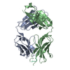
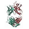
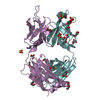
| Title | Unusual twinning in crystals of the CitS binding antibody Fab fragment f3p4 | ||||||
 Components Components |
| ||||||
 Keywords Keywords | IMMUNE SYSTEM / BINDING PROTEIN / RECOMBINANT FAB FRAGMENT / SYNTHETIC ANTIBODY LIBRARY (HUCAL) / PHAGE DISPLAY | ||||||
| Function / homology | Immunoglobulins / Immunoglobulin-like / Sandwich / Mainly Beta Function and homology information Function and homology information | ||||||
| Biological species | SYNTHETIC CONSTRUCT (others) | ||||||
| Method |  X-RAY DIFFRACTION / SYNCHROTRON / MOLECULAR REPLACEMENT / Resolution: 1.92 Å X-RAY DIFFRACTION / SYNCHROTRON / MOLECULAR REPLACEMENT / Resolution: 1.92 Å | ||||||
 Authors Authors | Frey, D. / Huber, T. / Plueckthun, A. / Gruetter, M.G. | ||||||
 Citation Citation | Journal: Acta Crystallogr.,Sect.D / Year: 2008 Title: Structure of the Recombinant Antibody Fab Fragment F3P4. Authors: Frey, D. / Huber, T. / Plueckthun, A. / Gruetter, M.G. | ||||||
| History |
| ||||||
| Remark 700 | SHEET THE SHEET STRUCTURE OF THIS MOLECULE IS BIFURCATED. IN ORDER TO REPRESENT THIS FEATURE IN ... SHEET THE SHEET STRUCTURE OF THIS MOLECULE IS BIFURCATED. IN ORDER TO REPRESENT THIS FEATURE IN THE SHEET RECORDS BELOW, TWO SHEETS ARE DEFINED. |
- Structure visualization
| Structure viewer | Molecule: MolmilJmol/JSmol |
|---|
- Downloads & links
-Download
| PDBx/mmCIF format | 2v7n.cif.gz | 332.2 KB | Display | PDBx/mmCIF format |
|---|---|---|---|---|
| PDB format | pdb2v7n.ent.gz | 270.3 KB | Display | PDB format |
| PDBx/mmJSON format | 2v7n.json.gz | Tree view | PDBx/mmJSON format | |
| Others |  Other downloads Other downloads |
-Validation report
| Arichive directory | https://data.pdbj.org/pub/pdb/validation_reports/v7/2v7nftp://data.pdbj.org/pub/pdb/validation_reports/v7/2v7n | HTTPS FTP |
|---|
-Related structure data
| Similar structure data |
|---|


-Links
 PDBj
PDBj

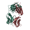
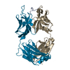
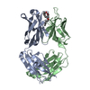
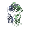
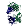
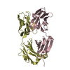
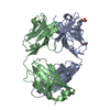
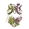
- Assembly
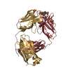
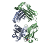
| Deposited unit | 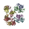
| ||||||||
|---|---|---|---|---|---|---|---|---|---|
| 1 | 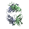
| ||||||||
| 2 | 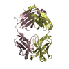
| ||||||||
| 3 | 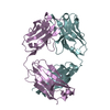
| ||||||||
| 4 | 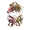
| ||||||||
| Unit cell |
|
-Components
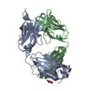
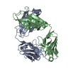
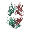
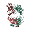
| #1: Antibody | Mass: 23382.799 Da / Num. of mol.: 4 / Fragment: FAB FRAGMENT, RESIDUES 1-255 Source method: isolated from a genetically manipulated source Details: SYNTHETIC GENE, DERIVED FROM PHAGE DISPLAY OF THE HUMAN COMBINATORIAL ANTIBODY LIBRARY (HUCAL) Source: (gene. exp.) SYNTHETIC CONSTRUCT (others) / Plasmid: PMX9 / Production host:  #2: Antibody | Mass: 24542.365 Da / Num. of mol.: 4 / Fragment: FAB FRAGMENT, RESIDUES 1-259 Source method: isolated from a genetically manipulated source Details: SYNTHETIC GENE, DERIVED FROM PHAGE DISPLAY OF THE HUMAN COMBINATORIAL ANTIBODY LIBRARY (HUCAL) Source: (gene. exp.) SYNTHETIC CONSTRUCT (others) / Plasmid: PMX9 / Production host: #3: Water | ChemComp-HOH / |  Mass: 18.015 Da / Num. of mol.: 540 / Source method: isolated from a natural source / Formula: H2O Mass: 18.015 Da / Num. of mol.: 540 / Source method: isolated from a natural source / Formula: H2OHas protein modification | Y | Sequence details | C-TERMINAL HISTIDINE TAG AT HEAVY CHAIN | |
|---|
-Experimental details
-Experiment
| Experiment | Method: X-RAY DIFFRACTION / Number of used crystals: 1 |
|---|
- Sample preparation
| Crystal | Density Matthews: 2.3 Å3/Da / Density % sol: 46.6 % / Description: NONE |
|---|---|
| Crystal grow | pH: 6.5 Details: 23% PEG4000, 50 MM LI2SO4, 50 MM NA2SO4, 50 MM N-(2ACETAMIDO) IMINODIACETIC ACID (ADA), PH 6.5 |
-Data collection
| Diffraction | Mean temperature: 100 K |
|---|---|
| Diffraction source | Source: SYNCHROTRON / Site: SLS  / Beamline: X06SA / Wavelength: 1 / Beamline: X06SA / Wavelength: 1 |
| Detector | Type: MARRESEARCH / Detector: CCD / Date: Oct 16, 2006 / Details: BENT MIRRORS |
| Radiation | Monochromator: SI(111) / Protocol: SINGLE WAVELENGTH / Monochromatic (M) / Laue (L): M / Scattering type: x-ray |
| Radiation wavelength | Wavelength: 1 Å / Relative weight: 1 |
| Reflection | Resolution: 1.9→31 Å / Num. obs: 151552 / % possible obs: 97.4 % / Observed criterion σ(I): 2 / Redundancy: 4.9 % / Rmerge(I) obs: 0.12 / Net I/σ(I): 10.2 |
| Reflection shell | Resolution: 1.9→1.92 Å / Redundancy: 3.9 % / Rmerge(I) obs: 0.37 / Mean I/σ(I) obs: 3.5 / % possible all: 83.3 |
- Processing
| Software |
| |||||||||||||||||||||||||||||||||
|---|---|---|---|---|---|---|---|---|---|---|---|---|---|---|---|---|---|---|---|---|---|---|---|---|---|---|---|---|---|---|---|---|---|---|
| Refinement | Method to determine structure: MOLECULAR REPLACEMENT / Resolution: 1.92→15 Å / Num. parameters: 53672 / Num. restraintsaints: 100475 / Cross valid method: FREE R-VALUE / σ(F): 0
| |||||||||||||||||||||||||||||||||
| Refine analyze | Num. disordered residues: 0 / Occupancy sum hydrogen: 0 / Occupancy sum non hydrogen: 13417 | |||||||||||||||||||||||||||||||||
| Refinement step | Cycle: LAST / Resolution: 1.92→15 Å
| |||||||||||||||||||||||||||||||||
| Refine LS restraints |
|
