 Movie
Movie Controller


+ Open data

- Basic information
| Entry | Database: PDB / ID: 1wcb | ||||||
|---|---|---|---|---|---|---|---|
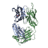
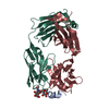
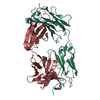
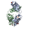
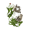
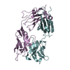
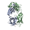
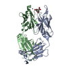
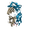
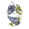
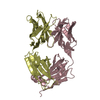
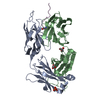
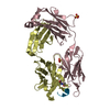
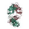
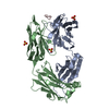
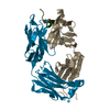
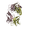
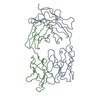
| Title | PLP-DEPENDENT CATALYTIC ANTIBODY 15A9 IN COMPLEX WITH ITS HAPTEN | ||||||
 Components Components |
| ||||||
 Keywords Keywords | IMMUNE SYSTEM / CATALYTIC ANTIBODY / TRANSAMINATION / PYRIDOXAL-PHOSPHATE / HAPTEN / PHOSPHOPYRIDOXYL-L-LYSINE | ||||||
| Function / homology | Immunoglobulins / Immunoglobulin-like / Sandwich / Mainly Beta / IODIDE ION / Chem-PE1 Function and homology information Function and homology information | ||||||
| Biological species |  | ||||||
| Method |  X-RAY DIFFRACTION / SYNCHROTRON / MOLECULAR REPLACEMENT / Resolution: 2.5 Å X-RAY DIFFRACTION / SYNCHROTRON / MOLECULAR REPLACEMENT / Resolution: 2.5 Å | ||||||
 Authors Authors | Golinelli-Pimpaneau, B. / Christen, P. | ||||||
 Citation Citation | Journal: J.Biol.Chem. / Year: 2006 Title: Structural Basis for D-Amino Acid Transamination by the Pyridoxal- 5' -Phosphate - Dependent Catalytic Antibody 15A9. Authors: Golinelli-Pimpaneau, B. / Luthi, C. / Christen, P. #1: Journal: J.Biol.Chem. / Year: 1997 Title: Monoclonal Antibodies Against N-Alpha-(5'-Phosphopyridoxyl)-L-Lysine. Authors: Gramatikova, S.I. / Christen, P. | ||||||
| History |
| ||||||
| Remark 700 | SHEET THE SHEET STRUCTURE OF THIS MOLECULE IS BIFURCATED. IN ORDER TO REPRESENT THIS FEATURE IN ... SHEET THE SHEET STRUCTURE OF THIS MOLECULE IS BIFURCATED. IN ORDER TO REPRESENT THIS FEATURE IN THE SHEET RECORDS BELOW, TWO SHEETS ARE DEFINED. |
- Structure visualization
| Structure viewer | Molecule: MolmilJmol/JSmol |
|---|
- Downloads & links
-Download
| PDBx/mmCIF format | 1wcb.cif.gz | 188.7 KB | Display | PDBx/mmCIF format |
|---|---|---|---|---|
| PDB format | pdb1wcb.ent.gz | 149.6 KB | Display | PDB format |
| PDBx/mmJSON format | 1wcb.json.gz | Tree view | PDBx/mmJSON format | |
| Others |  Other downloads Other downloads |
-Validation report
| Arichive directory | https://data.pdbj.org/pub/pdb/validation_reports/wc/1wcbftp://data.pdbj.org/pub/pdb/validation_reports/wc/1wcb | HTTPS FTP |
|---|
-Related structure data
| Related structure data |  2bmkC  1cloS C: citing same article ( S: Starting model for refinement |
|---|---|
| Similar structure data |


-Links
 PDBj
PDBj

- Assembly
| Deposited unit | 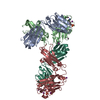
| ||||||||||||
|---|---|---|---|---|---|---|---|---|---|---|---|---|---|
| 1 | 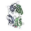
| ||||||||||||
| 2 | 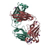
| ||||||||||||
| Unit cell |
| ||||||||||||
| Noncrystallographic symmetry (NCS) | NCS oper:
|
-Components
| #1: Antibody | Mass: 23355.787 Da / Num. of mol.: 2 Source method: isolated from a genetically manipulated source Source: (gene. exp.) #2: Antibody | Mass: 24501.449 Da / Num. of mol.: 2 Source method: isolated from a genetically manipulated source Source: (gene. exp.) #3: Chemical |   Type: L-peptide linking / Mass: 377.330 Da / Num. of mol.: 2 / Source method: obtained synthetically / Formula: C14H24N3O7P Type: L-peptide linking / Mass: 377.330 Da / Num. of mol.: 2 / Source method: obtained synthetically / Formula: C14H24N3O7P#4: Chemical | ChemComp-IOD /   Mass: 126.904 Da / Num. of mol.: 9 / Source method: obtained synthetically / Formula: I Mass: 126.904 Da / Num. of mol.: 9 / Source method: obtained synthetically / Formula: I#5: Water | ChemComp-HOH / |  Mass: 18.015 Da / Num. of mol.: 473 / Source method: isolated from a natural source / Formula: H2O Mass: 18.015 Da / Num. of mol.: 473 / Source method: isolated from a natural source / Formula: H2OHas protein modification | Y | |
|---|
-Experimental details
-Experiment
| Experiment | Method: X-RAY DIFFRACTION / Number of used crystals: 1 |
|---|
- Sample preparation
| Crystal | Density Matthews: 2.15 Å3/Da / Density % sol: 34.7 % |
|---|---|
| Crystal grow | Temperature: 289 K / Method: vapor diffusion, hanging drop / pH: 6 Details: 30% PEG 3350, 200MM SODIUM REMARK 280 IODIDE, 50MM SODIUM ACETATE, PH 6, VAPOR DIFFUSION, HANGING DROP, TEMPERATURE 289K |
-Data collection
| Diffraction | Mean temperature: 100 K |
|---|---|
| Diffraction source | Source: SYNCHROTRON / Site: ESRF  / Beamline: ID14-2 / Wavelength: 0.933 / Beamline: ID14-2 / Wavelength: 0.933 |
| Detector | Type: ADSC CCD / Detector: CCD / Date: Feb 24, 2003 |
| Radiation | Protocol: SINGLE WAVELENGTH / Monochromatic (M) / Laue (L): M / Scattering type: x-ray |
| Radiation wavelength | Wavelength: 0.933 Å / Relative weight: 1 |
| Reflection | Resolution: 2.5→25 Å / Num. obs: 28522 / % possible obs: 98.5 % / Observed criterion σ(I): 0 / Redundancy: 1.9 % / Biso Wilson estimate: 31.5 Å2 / Rmerge(I) obs: 0.08 / Net I/σ(I): 15.9 |
| Reflection shell | Resolution: 2.5→2.54 Å / Redundancy: 1.8 % / Rmerge(I) obs: 0.2 / Mean I/σ(I) obs: 5.1 / % possible all: 94.3 |
- Processing
| Software |
| ||||||||||||||||||||||||||||||||||||||||||||||||||||||||||||
|---|---|---|---|---|---|---|---|---|---|---|---|---|---|---|---|---|---|---|---|---|---|---|---|---|---|---|---|---|---|---|---|---|---|---|---|---|---|---|---|---|---|---|---|---|---|---|---|---|---|---|---|---|---|---|---|---|---|---|---|---|---|
| Refinement | Method to determine structure: MOLECULAR REPLACEMENT Starting model: PDB ENTRY 1CLO Resolution: 2.5→40 Å / Data cutoff high absF: 10000 / Cross valid method: THROUGHOUT / σ(F): 0 Stereochemistry target values: MAXIMUM LIKELIHOOD TARGET USING AMPLITUDES Details: THE FOLLOWING RESIDUES WERE NOT LOCATED IN THE ELECTRON DENSITY: ALA H 129, ALA H 130, ARG H 213, ASP H 214, ALA B 129, ALA B 130 THE SIDE-CHAINS OF THE FOLLOWING RESIDUES ARE POORLY DEFINED ...Details: THE FOLLOWING RESIDUES WERE NOT LOCATED IN THE ELECTRON DENSITY: ALA H 129, ALA H 130, ARG H 213, ASP H 214, ALA B 129, ALA B 130 THE SIDE-CHAINS OF THE FOLLOWING RESIDUES ARE POORLY DEFINED BY THE ELECTRON DENSITY AND WERE MODELLED AS ALANINE: SER L 56, LYS L 142, LYS H 43, ARG H 83, GLU H 100C, GLN H 131, SER H 134, SER H 158, GLN H 171, GLU H 191, LYS H 205, LYS H 209, GLN B 131, SER B 134, GLN B 171, LYS B 209
| ||||||||||||||||||||||||||||||||||||||||||||||||||||||||||||
| Solvent computation | Bsol: 36.4942 Å2 / ksol: 0.332759 e/Å3 | ||||||||||||||||||||||||||||||||||||||||||||||||||||||||||||
| Displacement parameters | Biso mean: 21.6 Å2
| ||||||||||||||||||||||||||||||||||||||||||||||||||||||||||||
| Refinement step | Cycle: LAST / Resolution: 2.5→40 Å
| ||||||||||||||||||||||||||||||||||||||||||||||||||||||||||||
| Refine LS restraints |
| ||||||||||||||||||||||||||||||||||||||||||||||||||||||||||||
| LS refinement shell | Resolution: 2.5→2.52 Å / Total num. of bins used: 50
| ||||||||||||||||||||||||||||||||||||||||||||||||||||||||||||
| Xplor file |
|
