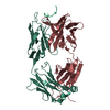
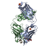
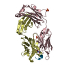
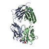
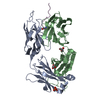
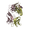
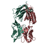
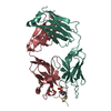
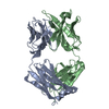
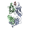
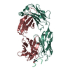
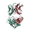
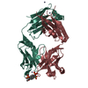
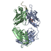
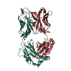
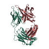
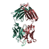
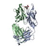
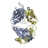
PROTEIN (IGG1FAB58.2ANTIBODY (LIGHTCHAIN)) / FAB 58.2
Mass: 23573.012 Da / Num. of mol.: 1 / Fragment: FAB FRAGMENT / Source method: isolated from a natural source / Source: (natural) Mus musculus (house mouse) / Strain: BALB/C / References: UniProt: P01666*PLUS
#2: Antibody
PROTEIN (IGG1FAB58.2ANTIBODY (HEAVYCHAIN)) / FAB 58.2
Mass: 24864.744 Da / Num. of mol.: 1 / Fragment: FAB FRAGMENT / Source method: isolated from a natural source / Source: (natural) Mus musculus (house mouse) / Strain: BALB/C
#3: Protein/peptide
PROTEIN (HIV-1GP120) / V3 LOOP
Mass: 1027.139 Da / Num. of mol.: 1 / Source method: obtained synthetically / Details: THIS MOLECULE WAS CHEMICALLY SYNTHESIZED
Type: XUONG-HAMLIN MULTIWIRE / Detector: AREA DETECTOR / Date: May 15, 1992
Radiation
Monochromator: GRAPHITE / Protocol: SINGLE WAVELENGTH / Monochromatic (M) / Laue (L): M / Scattering type: x-ray
Radiation wavelength
Wavelength: 1.5418 Å / Relative weight: 1
Reflection
Resolution: 2.8→24 Å / Num. obs: 12923 / % possible obs: 91.9 % / Redundancy: 3.4 % / Biso Wilson estimate: 35.1 Å2 / Rsym value: 0.076 / Net I/σ(I): 7.5
Reflection shell
Resolution: 2.8→3.02 Å / Redundancy: 1.99 % / Mean I/σ(I) obs: 1 / Rsym value: 0.288 / % possible all: 82.8
-
Processing
Software
Name
Version
Classification
UCSD-system
datacollection
UCSD-system
datareduction
MERLOT
phasing
X-PLOR
modelbuilding
X-PLOR
3.851
refinement
UCSD-system
datascaling
X-PLOR
phasing
Refinement
Method to determine structure: MOLECULAR REPLACEMENT Starting model: FAB 58.2 PORTION OF FAB 58.2/SER-LOOP PEPTIDE COMPLEX Resolution: 2.8→24 Å / Rfactor Rfree error: 0.012 / Data cutoff high absF: 10000000 / Data cutoff low absF: 0 / Isotropic thermal model: GROUP / Cross valid method: THROUGHOUT / σ(F): 0 / Details: BULK SOLVENT MODEL USED
Rfactor
Num. reflection
% reflection
Selection details
Rfree
0.305
615
4.8 %
RANDOM
Rwork
0.185
-
-
-
obs
0.185
12923
91.9 %
-
Displacement parameters
Biso mean: 26.7 Å2
Refine analyze
Free
Obs
Luzzati coordinate error
0.46 Å
0.28 Å
Luzzati d res low
-
5 Å
Luzzati sigma a
0.58 Å
0.42 Å
Refinement step
Cycle: LAST / Resolution: 2.8→24 Å
Protein
Nucleic acid
Ligand
Solvent
Total
Num. atoms
3485
0
0
1
3486
Refine LS restraints
Refine-ID
Type
Dev ideal
X-RAY DIFFRACTION
x_bond_d
0.012
X-RAY DIFFRACTION
x_bond_d_na
X-RAY DIFFRACTION
x_bond_d_prot
X-RAY DIFFRACTION
x_angle_d
X-RAY DIFFRACTION
x_angle_d_na
X-RAY DIFFRACTION
x_angle_d_prot
X-RAY DIFFRACTION
x_angle_deg
1.9
X-RAY DIFFRACTION
x_angle_deg_na
X-RAY DIFFRACTION
x_angle_deg_prot
X-RAY DIFFRACTION
x_dihedral_angle_d
28
X-RAY DIFFRACTION
x_dihedral_angle_d_na
X-RAY DIFFRACTION
x_dihedral_angle_d_prot
X-RAY DIFFRACTION
x_improper_angle_d
1.6
X-RAY DIFFRACTION
x_improper_angle_d_na
X-RAY DIFFRACTION
x_improper_angle_d_prot
X-RAY DIFFRACTION
x_mcbond_it
X-RAY DIFFRACTION
x_mcangle_it
X-RAY DIFFRACTION
x_scbond_it
X-RAY DIFFRACTION
x_scangle_it
LS refinement shell
Resolution: 2.8→3.02 Å / Rfactor Rfree error: 0.034 / Total num. of bins used: 5
Rfactor
Num. reflection
% reflection
Rfree
0.366
115
5 %
Rwork
0.276
2172
-
obs
-
-
82.8 %
Xplor file
Refine-ID
Serial no
Param file
Topol file
X-RAY DIFFRACTION
1
PARHCSDX.PRO
TOPHCSDX.PRO
X-RAY DIFFRACTION
2
PARAM11.WAT
TOPH11.WAT
+
About Yorodumi
-
News
-
Feb 9, 2022. New format data for meta-information of EMDB entries
New format data for meta-information of EMDB entries
Version 3 of the EMDB header file is now the official format.
The previous official version 1.9 will be removed from the archive.
In the structure databanks used in Yorodumi, some data are registered as the other names, "COVID-19 virus" and "2019-nCoV". Here are the details of the virus and the list of structure data.
Jan 31, 2019. EMDB accession codes are about to change! (news from PDBe EMDB page)
EMDB accession codes are about to change! (news from PDBe EMDB page)
The allocation of 4 digits for EMDB accession codes will soon come to an end. Whilst these codes will remain in use, new EMDB accession codes will include an additional digit and will expand incrementally as the available range of codes is exhausted. The current 4-digit format prefixed with “EMD-” (i.e. EMD-XXXX) will advance to a 5-digit format (i.e. EMD-XXXXX), and so on. It is currently estimated that the 4-digit codes will be depleted around Spring 2019, at which point the 5-digit format will come into force.
The EM Navigator/Yorodumi systems omit the EMD- prefix.
Related info.:Q: What is EMD? / ID/Accession-code notation in Yorodumi/EM Navigator
Yorodumi is a browser for structure data from EMDB, PDB, SASBDB, etc.
This page is also the successor to EM Navigator detail page, and also detail information page/front-end page for Omokage search.
The word "yorodu" (or yorozu) is an old Japanese word meaning "ten thousand". "mi" (miru) is to see.
Related info.:EMDB / PDB / SASBDB / Comparison of 3 databanks / Yorodumi Search / Aug 31, 2016. New EM Navigator & Yorodumi / Yorodumi Papers / Jmol/JSmol / Function and homology information / Changes in new EM Navigator and Yorodumi
 Movie
Movie Controller

 Yorodumi
 Open data
 Basic information
 Components
 Keywords
 Function and homology information
 X-RAY DIFFRACTION /
  Authors
 Citation
 Structure visualization
 Downloads & links
 Other downloads
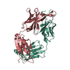

 PDBj
PDBj


 Assembly
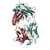
 Mass: 99.131 Da / Num. of mol.: 1 / Source method: obtained synthetically / Formula: C5H9NO
 Mass: 18.015 Da / Num. of mol.: 1 / Source method: isolated from a natural source / Formula: H2O
 Sample preparation
 Processing