 Movie
Movie Controller

[English] 日本語
 Yorodumi
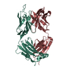
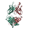
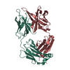
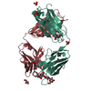
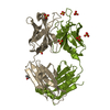
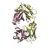
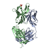
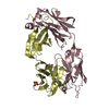
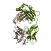
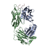
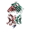
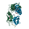
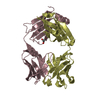
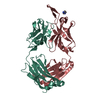
- PDB-6gxx: Fab fragment of an antibody selective for alpha-1-antitrypsin in ... -
+ Open data

- Basic information
| Entry | Database: PDB / ID: 6gxx | ||||||||||||||||||
|---|---|---|---|---|---|---|---|---|---|---|---|---|---|---|---|---|---|---|---|
| Title | Fab fragment of an antibody selective for alpha-1-antitrypsin in the native conformation | ||||||||||||||||||
 Components Components |
| ||||||||||||||||||
 Keywords Keywords | PROTEIN BINDING / Antibody fragment / Antitrypsin binding / Diagnostic / Monoclonal / conformationally-selective | ||||||||||||||||||
| Function / homology | Immunoglobulins / Immunoglobulin-like / Sandwich / Mainly Beta Function and homology information Function and homology information | ||||||||||||||||||
| Biological species |  | ||||||||||||||||||
| Method |  X-RAY DIFFRACTION / SYNCHROTRON / MOLECULAR REPLACEMENT / Resolution: 1.85 Å X-RAY DIFFRACTION / SYNCHROTRON / MOLECULAR REPLACEMENT / Resolution: 1.85 Å | ||||||||||||||||||
 Authors Authors | Elliston, E.L.K. / Miranda, E. / Perez, J. / Lomas, D.A. / Irving, J.A. | ||||||||||||||||||
| Funding support |  United Kingdom, United Kingdom,  United States, 5items United States, 5items
| ||||||||||||||||||
 Citation Citation | Journal: To Be Published Title: Characterisation of a monoclonal antibody conformationally-selective for native alpha-1-antitrypsin Authors: Elliston, E.L.K. / Miranda, E. / Perez, J. / Irving, J.A. / Lomas, D.A. | ||||||||||||||||||
| History |
|
- Structure visualization
| Structure viewer | Molecule: MolmilJmol/JSmol |
|---|
- Downloads & links
-Download
| PDBx/mmCIF format | 6gxx.cif.gz | 115.1 KB | Display | PDBx/mmCIF format |
|---|---|---|---|---|
| PDB format | pdb6gxx.ent.gz | 82.8 KB | Display | PDB format |
| PDBx/mmJSON format | 6gxx.json.gz | Tree view | PDBx/mmJSON format | |
| Others |  Other downloads Other downloads |
-Validation report
| Arichive directory | https://data.pdbj.org/pub/pdb/validation_reports/gx/6gxxftp://data.pdbj.org/pub/pdb/validation_reports/gx/6gxx | HTTPS FTP |
|---|
-Related structure data
| Related structure data |  6hx4C  1mf2S S: Starting model for refinement C: citing same article ( |
|---|---|
| Similar structure data |


-Links
 PDBj
PDBj

- Assembly
| Deposited unit | 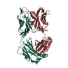
| ||||||||||||||||||
|---|---|---|---|---|---|---|---|---|---|---|---|---|---|---|---|---|---|---|---|
| 1 |
| ||||||||||||||||||
| Unit cell |
| ||||||||||||||||||
| Components on special symmetry positions |
|
-Components
| #1: Antibody | Mass: 24161.053 Da / Num. of mol.: 1 / Source method: isolated from a natural source / Source: (natural) |
|---|---|
| #2: Antibody | Mass: 23859.277 Da / Num. of mol.: 1 / Source method: isolated from a natural source / Source: (natural) |
| #3: Chemical | ChemComp-MG /   Mass: 24.305 Da / Num. of mol.: 1 / Source method: obtained synthetically / Formula: Mg Mass: 24.305 Da / Num. of mol.: 1 / Source method: obtained synthetically / Formula: Mg |
| #4: Water | ChemComp-HOH /  Mass: 18.015 Da / Num. of mol.: 584 / Source method: isolated from a natural source / Formula: H2O Mass: 18.015 Da / Num. of mol.: 584 / Source method: isolated from a natural source / Formula: H2O |
| Has protein modification | Y |
-Experimental details
-Experiment
| Experiment | Method: X-RAY DIFFRACTION / Number of used crystals: 1 |
|---|
- Sample preparation
| Crystal | Density Matthews: 2.13 Å3/Da / Density % sol: 42.21 % / Description: Tabular |
|---|---|
| Crystal grow | Temperature: 289 K / Method: vapor diffusion, hanging drop / pH: 6.5 / Details: 20% PEG 3350, 0.1M Hepes, 0.05M magnesium chloride |
-Data collection
| Diffraction | Mean temperature: 100 K / Ambient temp details: Cryostream | ||||||||||||||||||||||||
|---|---|---|---|---|---|---|---|---|---|---|---|---|---|---|---|---|---|---|---|---|---|---|---|---|---|
| Diffraction source | Source: SYNCHROTRON / Site: Diamond / Beamline: I03 / Wavelength: 0.99998 Å | ||||||||||||||||||||||||
| Detector | Type: DECTRIS PILATUS3 6M / Detector: PIXEL / Date: Feb 11, 2018 | ||||||||||||||||||||||||
| Radiation | Monochromator: Double crystal Si(111) / Protocol: SINGLE WAVELENGTH / Monochromatic (M) / Laue (L): M / Scattering type: x-ray | ||||||||||||||||||||||||
| Radiation wavelength | Wavelength: 0.99998 Å / Relative weight: 1 | ||||||||||||||||||||||||
| Reflection | Resolution: 1.85→29.81 Å / Num. obs: 34148 / % possible obs: 94.9 % / Redundancy: 12.6 % / Biso Wilson estimate: 19.06 Å2 / CC1/2: 0.998 / Rmerge(I) obs: 0.133 / Rpim(I) all: 0.039 / Rrim(I) all: 0.139 / Net I/σ(I): 15.3 | ||||||||||||||||||||||||
| Reflection shell | Diffraction-ID: 1
|
- Processing
| Software |
| |||||||||||||||||||||||||||||||||||||||||||||||||||||||||||||||||||||||||||||||||||||||||||
|---|---|---|---|---|---|---|---|---|---|---|---|---|---|---|---|---|---|---|---|---|---|---|---|---|---|---|---|---|---|---|---|---|---|---|---|---|---|---|---|---|---|---|---|---|---|---|---|---|---|---|---|---|---|---|---|---|---|---|---|---|---|---|---|---|---|---|---|---|---|---|---|---|---|---|---|---|---|---|---|---|---|---|---|---|---|---|---|---|---|---|---|---|
| Refinement | Method to determine structure: MOLECULAR REPLACEMENT Starting model: 1mf2 Resolution: 1.85→27.08 Å / SU ML: 0.2351 / Cross valid method: FREE R-VALUE / σ(F): 1.34 / Phase error: 22.4849
| |||||||||||||||||||||||||||||||||||||||||||||||||||||||||||||||||||||||||||||||||||||||||||
| Solvent computation | Shrinkage radii: 0.8 Å / VDW probe radii: 1 Å | |||||||||||||||||||||||||||||||||||||||||||||||||||||||||||||||||||||||||||||||||||||||||||
| Displacement parameters | Biso mean: 19.96 Å2 | |||||||||||||||||||||||||||||||||||||||||||||||||||||||||||||||||||||||||||||||||||||||||||
| Refinement step | Cycle: LAST / Resolution: 1.85→27.08 Å
| |||||||||||||||||||||||||||||||||||||||||||||||||||||||||||||||||||||||||||||||||||||||||||
| Refine LS restraints |
| |||||||||||||||||||||||||||||||||||||||||||||||||||||||||||||||||||||||||||||||||||||||||||
| LS refinement shell |
|
