 Movie
Movie Controller

[English] 日本語
 Yorodumi
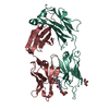
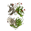
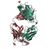
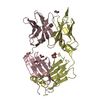
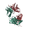
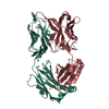
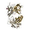
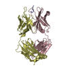
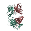
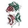
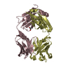
- PDB-2vxu: Crystal structure of murine reference antibody 125-2H Fab fragment -
+ Open data

- Basic information
| Entry | Database: PDB / ID: 2vxu | ||||||
|---|---|---|---|---|---|---|---|
| Title | Crystal structure of murine reference antibody 125-2H Fab fragment | ||||||
 Components Components | (MURINE IGG 125-2H) x 2 | ||||||
 Keywords Keywords | IMMUNE SYSTEM / FAB / IL-18 / AUTOIMMUNITY / TH1/TH2 CELLS | ||||||
| Function / homology | Immunoglobulins / Immunoglobulin-like / Sandwich / Mainly Beta Function and homology information Function and homology information | ||||||
| Biological species |  | ||||||
| Method |  X-RAY DIFFRACTION / SYNCHROTRON / MOLECULAR REPLACEMENT / Resolution: 2.36 Å X-RAY DIFFRACTION / SYNCHROTRON / MOLECULAR REPLACEMENT / Resolution: 2.36 Å | ||||||
 Authors Authors | Argiriadi, M.A. / Xiang, T. / Wu, C. / Ghayur, T. / Borhani, D.W. | ||||||
 Citation Citation | Journal: J.Biol.Chem. / Year: 2009 Title: Unusual Water-Mediated Antigenic Recognition of the Proinflammatory Cytokine Interleukin-18. Authors: Argiriadi, M.A. / Xiang, T. / Wu, C. / Ghayur, T. / Borhani, D.W. | ||||||
| History |
| ||||||
| Remark 700 | SHEET THE SHEET STRUCTURE OF THIS MOLECULE IS BIFURCATED. IN ORDER TO REPRESENT THIS FEATURE IN ... SHEET THE SHEET STRUCTURE OF THIS MOLECULE IS BIFURCATED. IN ORDER TO REPRESENT THIS FEATURE IN THE SHEET RECORDS BELOW, TWO SHEETS ARE DEFINED. |
- Structure visualization
| Structure viewer | Molecule: MolmilJmol/JSmol |
|---|
- Downloads & links
-Download
| PDBx/mmCIF format | 2vxu.cif.gz | 185.1 KB | Display | PDBx/mmCIF format |
|---|---|---|---|---|
| PDB format | pdb2vxu.ent.gz | 148.1 KB | Display | PDB format |
| PDBx/mmJSON format | 2vxu.json.gz | Tree view | PDBx/mmJSON format | |
| Others |  Other downloads Other downloads |
-Validation report
| Arichive directory | https://data.pdbj.org/pub/pdb/validation_reports/vx/2vxuftp://data.pdbj.org/pub/pdb/validation_reports/vx/2vxu | HTTPS FTP |
|---|
-Related structure data
| Related structure data |  2vxtC 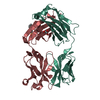 2vxvC 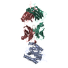 1fnsS C: citing same article ( S: Starting model for refinement |
|---|---|
| Similar structure data |


-Links
 PDBj
PDBj

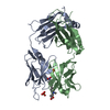
- Assembly
| Deposited unit | 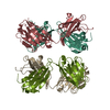
| ||||||||||||
|---|---|---|---|---|---|---|---|---|---|---|---|---|---|
| 1 | 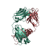
| ||||||||||||
| 2 | 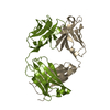
| ||||||||||||
| Unit cell |
| ||||||||||||
| Noncrystallographic symmetry (NCS) | NCS oper:
|
-Components
| #1: Antibody | Mass: 23260.008 Da / Num. of mol.: 2 Source method: isolated from a genetically manipulated source Source: (gene. exp.) #2: Antibody | Mass: 23625.969 Da / Num. of mol.: 2 Source method: isolated from a genetically manipulated source Source: (gene. exp.) #3: Water | ChemComp-HOH / |  Mass: 18.015 Da / Num. of mol.: 488 / Source method: isolated from a natural source / Formula: H2O Mass: 18.015 Da / Num. of mol.: 488 / Source method: isolated from a natural source / Formula: H2OHas protein modification | Y | |
|---|
-Experimental details
-Experiment
| Experiment | Method: X-RAY DIFFRACTION / Number of used crystals: 1 |
|---|
- Sample preparation
| Crystal | Density Matthews: 2.7 Å3/Da / Density % sol: 54.3 % / Description: NONE |
|---|---|
| Crystal grow | Temperature: 277 K / pH: 7.5 Details: FAB (13 MG/ML, 2 MICROLITERS [UL]) WAS MIXED WITH 2 UL OF RESERVOIR (10% POLYETHYLENEGLYCOL (PEG) 6000, 100 MM HEPES, PH 7.5, 5% 2, 4-METHYLPENTANEDIOL) AND SUSPENDED OVER THE RESERVOIR AT ...Details: FAB (13 MG/ML, 2 MICROLITERS [UL]) WAS MIXED WITH 2 UL OF RESERVOIR (10% POLYETHYLENEGLYCOL (PEG) 6000, 100 MM HEPES, PH 7.5, 5% 2, 4-METHYLPENTANEDIOL) AND SUSPENDED OVER THE RESERVOIR AT 277 K. ROD-LIKE CRYSTALS APPEARED WITHIN ONE DAY. |
-Data collection
| Diffraction | Mean temperature: 100 K |
|---|---|
| Diffraction source | Source: SYNCHROTRON / Site: APS  / Beamline: 17-ID / Wavelength: 1 / Beamline: 17-ID / Wavelength: 1 |
| Detector | Type: MARRESEARCH / Detector: CCD / Date: Mar 20, 2003 / Details: MIRROR |
| Radiation | Monochromator: SI 111 / Protocol: SINGLE WAVELENGTH / Monochromatic (M) / Laue (L): M / Scattering type: x-ray |
| Radiation wavelength | Wavelength: 1 Å / Relative weight: 1 |
| Reflection | Resolution: 2.33→17.5 Å / Num. obs: 41229 / % possible obs: 94 % / Observed criterion σ(I): 2 / Redundancy: 5.2 % / Biso Wilson estimate: 41 Å2 / Rmerge(I) obs: 0.05 / Net I/σ(I): 18.9 |
| Reflection shell | Resolution: 2.33→2.39 Å / Redundancy: 2.4 % / Rmerge(I) obs: 0.24 / Mean I/σ(I) obs: 4.9 / % possible all: 25.2 |
- Processing
| Software |
| ||||||||||||||||||||||||||||||||||||||||||||||||||||||||||||||||||||||||||||||||||||||||||||||||||||||||||||||||||||||||||||||||||||||||||||||||||||||||||||||||||||||||||||||||||||||
|---|---|---|---|---|---|---|---|---|---|---|---|---|---|---|---|---|---|---|---|---|---|---|---|---|---|---|---|---|---|---|---|---|---|---|---|---|---|---|---|---|---|---|---|---|---|---|---|---|---|---|---|---|---|---|---|---|---|---|---|---|---|---|---|---|---|---|---|---|---|---|---|---|---|---|---|---|---|---|---|---|---|---|---|---|---|---|---|---|---|---|---|---|---|---|---|---|---|---|---|---|---|---|---|---|---|---|---|---|---|---|---|---|---|---|---|---|---|---|---|---|---|---|---|---|---|---|---|---|---|---|---|---|---|---|---|---|---|---|---|---|---|---|---|---|---|---|---|---|---|---|---|---|---|---|---|---|---|---|---|---|---|---|---|---|---|---|---|---|---|---|---|---|---|---|---|---|---|---|---|---|---|---|---|
| Refinement | Method to determine structure: MOLECULAR REPLACEMENT Starting model: PDB ENTRY 1FNS Resolution: 2.36→17.42 Å / Cor.coef. Fo:Fc: 0.946 / Cor.coef. Fo:Fc free: 0.916 / SU B: 13.513 / SU ML: 0.174 / TLS residual ADP flag: LIKELY RESIDUAL / Cross valid method: THROUGHOUT / ESU R: 0.371 / ESU R Free: 0.251 / Stereochemistry target values: MAXIMUM LIKELIHOOD Details: HYDROGENS HAVE BEEN ADDED IN THE RIDING POSITIONS. MOLECULE A (CHAINS H AND L) AND A CRYSTALLOGRAPHIC SYMMETRY MATE OF MOLECULE B (CHAINS I AND M) ARE RELATED BY TRANSLATIONAL PSEUDO- ...Details: HYDROGENS HAVE BEEN ADDED IN THE RIDING POSITIONS. MOLECULE A (CHAINS H AND L) AND A CRYSTALLOGRAPHIC SYMMETRY MATE OF MOLECULE B (CHAINS I AND M) ARE RELATED BY TRANSLATIONAL PSEUDO-SYMMETRY. MOLECULE A IS TRANSFORMED ONTO MOLECULE B_SYM BY ROTATION OF 4.4 DEGREES ABOUT AN AXIS WITH DIRECTION COSINES (-0.6574, 0.3326, 0. 6762) FOLLOWED BY TRANSLATION OF (0.7249, 46.9819, 53.2092) ANGSTROM UNITS.
| ||||||||||||||||||||||||||||||||||||||||||||||||||||||||||||||||||||||||||||||||||||||||||||||||||||||||||||||||||||||||||||||||||||||||||||||||||||||||||||||||||||||||||||||||||||||
| Solvent computation | Ion probe radii: 0.8 Å / Shrinkage radii: 0.8 Å / VDW probe radii: 1.4 Å / Solvent model: BABINET MODEL WITH MASK | ||||||||||||||||||||||||||||||||||||||||||||||||||||||||||||||||||||||||||||||||||||||||||||||||||||||||||||||||||||||||||||||||||||||||||||||||||||||||||||||||||||||||||||||||||||||
| Displacement parameters | Biso mean: 49.19 Å2
| ||||||||||||||||||||||||||||||||||||||||||||||||||||||||||||||||||||||||||||||||||||||||||||||||||||||||||||||||||||||||||||||||||||||||||||||||||||||||||||||||||||||||||||||||||||||
| Refinement step | Cycle: LAST / Resolution: 2.36→17.42 Å
| ||||||||||||||||||||||||||||||||||||||||||||||||||||||||||||||||||||||||||||||||||||||||||||||||||||||||||||||||||||||||||||||||||||||||||||||||||||||||||||||||||||||||||||||||||||||
| Refine LS restraints |
|