 Movie
Movie Controller

[English] 日本語
 Yorodumi
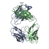
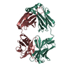
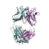
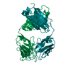
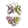
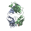
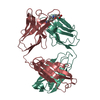
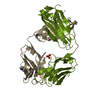
- PDB-6kva: Structure of anti-hCXCR2 abN48-2 in complex with its CXCR2 epitope -
+ Open data

- Basic information
| Entry | Database: PDB / ID: 6kva | ||||||
|---|---|---|---|---|---|---|---|
| Title | Structure of anti-hCXCR2 abN48-2 in complex with its CXCR2 epitope | ||||||
 Components Components |
| ||||||
 Keywords Keywords | IMMUNE SYSTEM / Antibody-Antigen Complex | ||||||
| Function / homology |  Function and homology information Function and homology informationinterleukin-8-mediated signaling pathway / interleukin-8 receptor activity / mast cell granule / interleukin-8 binding / C-X-C chemokine receptor activity / neutrophil activation / C-C chemokine receptor activity / C-C chemokine binding / Chemokine receptors bind chemokines / dendritic cell chemotaxis ...interleukin-8-mediated signaling pathway / interleukin-8 receptor activity / mast cell granule / interleukin-8 binding / C-X-C chemokine receptor activity / neutrophil activation / C-C chemokine receptor activity / C-C chemokine binding / Chemokine receptors bind chemokines / dendritic cell chemotaxis / cellular defense response / neutrophil chemotaxis / secretory granule membrane / calcium-mediated signaling / receptor internalization / G protein-coupled receptor activity / chemotaxis / mitotic spindle / microtubule cytoskeleton / positive regulation of cytosolic calcium ion concentration / G alpha (i) signalling events / phospholipase C-activating G protein-coupled receptor signaling pathway / cell surface receptor signaling pathway / immune response / inflammatory response / external side of plasma membrane / positive regulation of cell population proliferation / Neutrophil degranulation / negative regulation of apoptotic process / cell surface / signal transduction / nucleoplasm / membrane / plasma membrane Similarity search - Function | ||||||
| Biological species |  Homo sapiens (human) Homo sapiens (human) | ||||||
| Method |  X-RAY DIFFRACTION / SYNCHROTRON / MOLECULAR REPLACEMENT / Resolution: 2.2 Å X-RAY DIFFRACTION / SYNCHROTRON / MOLECULAR REPLACEMENT / Resolution: 2.2 Å | ||||||
 Authors Authors | Xiang, J.C. / Yan, L. / Yang, B. / Wilson, I.A. | ||||||
| Funding support |  China, 1items China, 1items
| ||||||
 Citation Citation | Journal: Nat Commun / Year: 2021 Title: Selection of a picomolar antibody that targets CXCR2-mediated neutrophil activation and alleviates EAE symptoms. Authors: Shi, X. / Wan, Y. / Wang, N. / Xiang, J. / Wang, T. / Yang, X. / Wang, J. / Dong, X. / Dong, L. / Yan, L. / Li, Y. / Liu, L. / Hou, S. / Zhong, Z. / Wilson, I.A. / Yang, B. / Yang, G. / Lerner, R.A. | ||||||
| History |
|
- Structure visualization
| Structure viewer | Molecule: MolmilJmol/JSmol |
|---|
- Downloads & links
-Download
| PDBx/mmCIF format | 6kva.cif.gz | 185.2 KB | Display | PDBx/mmCIF format |
|---|---|---|---|---|
| PDB format | pdb6kva.ent.gz | 145.7 KB | Display | PDB format |
| PDBx/mmJSON format | 6kva.json.gz | Tree view | PDBx/mmJSON format | |
| Others |  Other downloads Other downloads |
-Validation report
| Arichive directory | https://data.pdbj.org/pub/pdb/validation_reports/kv/6kvaftp://data.pdbj.org/pub/pdb/validation_reports/kv/6kva | HTTPS FTP |
|---|
-Related structure data
| Related structure data |  6kvfC  4xcnS S: Starting model for refinement C: citing same article ( |
|---|---|
| Similar structure data |


-Links
 PDBj
PDBj


- Assembly
| Deposited unit | 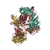
| ||||||||
|---|---|---|---|---|---|---|---|---|---|
| 1 | 
| ||||||||
| 2 | 
| ||||||||
| Unit cell |
|
-Components
| #1: Antibody | Mass: 24120.109 Da / Num. of mol.: 2 Source method: isolated from a genetically manipulated source Source: (gene. exp.) Homo sapiens (human) / Production host: Homo sapiens (human)#2: Antibody | Mass: 23646.229 Da / Num. of mol.: 2 Source method: isolated from a genetically manipulated source Source: (gene. exp.) Homo sapiens (human) / Production host: Homo sapiens (human)#3: Protein/peptide | Mass: 1375.373 Da / Num. of mol.: 2 / Source method: obtained synthetically / Source: (synth.) Homo sapiens (human) / References: UniProt: P25025#4: Chemical |   Mass: 62.068 Da / Num. of mol.: 2 / Source method: obtained synthetically / Formula: C2H6O2 Mass: 62.068 Da / Num. of mol.: 2 / Source method: obtained synthetically / Formula: C2H6O2#5: Water | ChemComp-HOH / |  Mass: 18.015 Da / Num. of mol.: 428 / Source method: isolated from a natural source / Formula: H2O Mass: 18.015 Da / Num. of mol.: 428 / Source method: isolated from a natural source / Formula: H2OHas ligand of interest | N | Has protein modification | Y | |
|---|
-Experimental details
-Experiment
| Experiment | Method: X-RAY DIFFRACTION / Number of used crystals: 1 |
|---|
- Sample preparation
| Crystal | Density Matthews: 2.18 Å3/Da / Density % sol: 43.47 % |
|---|---|
| Crystal grow | Temperature: 293 K / Method: vapor diffusion, hanging drop / pH: 7.5 Details: 0.1M HEPES pH 7.5, 25% w/v Polyethylene glycol 3350 PH range: 7.0-8.0 |
-Data collection
| Diffraction | Mean temperature: 100 K / Serial crystal experiment: N |
|---|---|
| Diffraction source | Source: SYNCHROTRON / Site: SSRF / Beamline: BL19U1 / Wavelength: 0.978 Å |
| Detector | Type: DECTRIS PILATUS3 S 6M / Detector: PIXEL / Date: Dec 15, 2017 |
| Radiation | Protocol: SINGLE WAVELENGTH / Monochromatic (M) / Laue (L): M / Scattering type: x-ray |
| Radiation wavelength | Wavelength: 0.978 Å / Relative weight: 1 |
| Reflection | Resolution: 2.2→50 Å / Num. obs: 42603 / % possible obs: 99.2 % / Redundancy: 4.7 % / Biso Wilson estimate: 31 Å2 / CC1/2: 1 / Net I/σ(I): 12.9 |
| Reflection shell | Resolution: 2.2→2.28 Å / Redundancy: 4 % / Num. unique obs: 2473 / CC1/2: 0.83 / % possible all: 94.2 |
- Processing
| Software |
| ||||||||||||||||||||||||||||||||||||||||||||||||||||||||||||
|---|---|---|---|---|---|---|---|---|---|---|---|---|---|---|---|---|---|---|---|---|---|---|---|---|---|---|---|---|---|---|---|---|---|---|---|---|---|---|---|---|---|---|---|---|---|---|---|---|---|---|---|---|---|---|---|---|---|---|---|---|---|
| Refinement | Method to determine structure: MOLECULAR REPLACEMENT Starting model: 4XCN Resolution: 2.2→48.56 Å / Cor.coef. Fo:Fc: 0.95 / Cor.coef. Fo:Fc free: 0.925 / Cross valid method: THROUGHOUT / σ(F): 0 / ESU R: 0.328 / ESU R Free: 0.219 Details: HYDROGENS HAVE BEEN ADDED IN THE RIDING POSITIONS U VALUES : REFINED INDIVIDUALLY
| ||||||||||||||||||||||||||||||||||||||||||||||||||||||||||||
| Solvent computation | Ion probe radii: 0.8 Å / Shrinkage radii: 0.8 Å / VDW probe radii: 1.2 Å | ||||||||||||||||||||||||||||||||||||||||||||||||||||||||||||
| Displacement parameters | Biso max: 75.96 Å2 / Biso mean: 32 Å2 / Biso min: 16.82 Å2
| ||||||||||||||||||||||||||||||||||||||||||||||||||||||||||||
| Refinement step | Cycle: final / Resolution: 2.2→48.56 Å
| ||||||||||||||||||||||||||||||||||||||||||||||||||||||||||||
| Refine LS restraints |
| ||||||||||||||||||||||||||||||||||||||||||||||||||||||||||||
| LS refinement shell | Resolution: 2.2→2.253 Å / Rfactor Rfree error: 0
|
