- PDB-1fns: CRYSTAL STRUCTURE OF THE VON WILLEBRAND FACTOR (VWF) A1 DOMAIN I5... -
+
Open data
ID or keywords:
Loading...
-
Basic information
Entry
Database: PDB / ID: 1fns
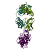
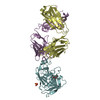
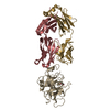
Title
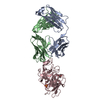
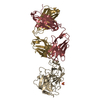
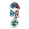
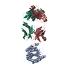
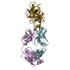
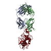
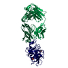
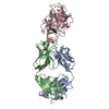
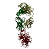
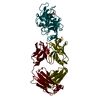
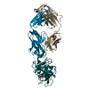
CRYSTAL STRUCTURE OF THE VON WILLEBRAND FACTOR (VWF) A1 DOMAIN I546V MUTANT IN COMPLEX WITH THE FUNCTION BLOCKING FAB NMC4
Components
(IMMUNOGLOBULIN NMC-4 IGG1) x 2
VON WILLEBRAND FACTOR
Keywords
IMMUNE SYSTEM / VON WILLEBRAND FACTOR / GLYCOPROTEIN IBA (A:ALPHA) BINDING / COMPLEX (WILLEBRAND-IMMUNOGLOBULIN) / BLOOD COAGULATION TYPE 2B VON WILLEBRAND DISEASE
Function / homology
Function and homology information
Defective VWF binding to collagen type I / Enhanced cleavage of VWF variant by ADAMTS13 / Defective VWF cleavage by ADAMTS13 variant / Defective F8 binding to von Willebrand factor / Enhanced binding of GP1BA variant to VWF multimer:collagen / Defective binding of VWF variant to GPIb:IX:V / Weibel-Palade body / hemostasis / platelet alpha granule / Platelet Adhesion to exposed collagen ...Defective VWF binding to collagen type I / Enhanced cleavage of VWF variant by ADAMTS13 / Defective VWF cleavage by ADAMTS13 variant / Defective F8 binding to von Willebrand factor / Enhanced binding of GP1BA variant to VWF multimer:collagen / Defective binding of VWF variant to GPIb:IX:V / Weibel-Palade body / hemostasis / platelet alpha granule / Platelet Adhesion to exposed collagen / alpha-beta T cell receptor complex / IgG immunoglobulin complex / extracellular matrix structural constituent / GP1b-IX-V activation signalling / p130Cas linkage to MAPK signaling for integrins / Defective F8 cleavage by thrombin / Platelet Aggregation (Plug Formation) / cell-substrate adhesion / GRB2:SOS provides linkage to MAPK signaling for Integrins / positive regulation of intracellular signal transduction / immunoglobulin binding / Integrin cell surface interactions / collagen binding / : / Integrin signaling / platelet alpha granule lumen / B cell differentiation / Signaling by high-kinase activity BRAF mutants / MAP2K and MAPK activation / platelet activation / response to wounding / integrin binding / blood coagulation / Signaling by RAF1 mutants / Signaling by moderate kinase activity BRAF mutants / Paradoxical activation of RAF signaling by kinase inactive BRAF / Signaling downstream of RAS mutants / Signaling by BRAF and RAF1 fusions / Platelet degranulation / protein-folding chaperone binding / extracellular matrix / protease binding / adaptive immune response / cell adhesion / endoplasmic reticulum / : / extracellular exosome / extracellular region / identical protein binding / plasma membrane Similarity search - Function
von Willebrand factor, VWA N-terminal domain / Von Willebrand factor / VWA N-terminal / Von Willebrand factor-like domain / : / Otogelin-like/Mucin TIL domain / C8 domain / Uncharacterised domain, cysteine-rich / : / C8 ...von Willebrand factor, VWA N-terminal domain / Von Willebrand factor / VWA N-terminal / Von Willebrand factor-like domain / : / Otogelin-like/Mucin TIL domain / C8 domain / Uncharacterised domain, cysteine-rich / : / C8 / von Willebrand factor, type D domain / von Willebrand factor type D domain / VWFD domain profile. / von Willebrand factor (vWF) type D domain / Trypsin Inhibitor-like, cysteine rich domain / Serine protease inhibitor-like superfamily / Trypsin Inhibitor like cysteine rich domain / C-terminal cystine knot signature. / C-terminal cystine knot domain profile. / von Willebrand factor (vWF) type C domain / von Willebrand factor type C domain / Cystine knot, C-terminal / C-terminal cystine knot-like domain (CTCK) / VWFC domain signature. / VWFC domain profile. / von Willebrand factor (vWF) type C domain / VWFC domain / von Willebrand factor, type A domain / von Willebrand factor type A domain / VWFA domain profile. / von Willebrand factor (vWF) type A domain / : / von Willebrand factor, type A / von Willebrand factor A-like domain superfamily / Immunoglobulin/major histocompatibility complex, conserved site / Immunoglobulins and major histocompatibility complex proteins signature. / Immunoglobulin C-Type / Immunoglobulin C1-set / Immunoglobulin C1-set domain / Ig-like domain profile. / Immunoglobulin-like domain / Immunoglobulin-like domain superfamily / Immunoglobulin-like fold / Immunoglobulins / Immunoglobulin-like / Sandwich / Rossmann fold / 3-Layer(aba) Sandwich / Mainly Beta / Alpha Beta Similarity search - Domain/homology
Immunoglobulin kappa constant / Ig gamma-1 chain C region secreted form / von Willebrand factor Similarity search - Component
In the structure databanks used in Yorodumi, some data are registered as the other names, "COVID-19 virus" and "2019-nCoV". Here are the details of the virus and the list of structure data.
Jan 31, 2019. EMDB accession codes are about to change! (news from PDBe EMDB page)
EMDB accession codes are about to change! (news from PDBe EMDB page)
The allocation of 4 digits for EMDB accession codes will soon come to an end. Whilst these codes will remain in use, new EMDB accession codes will include an additional digit and will expand incrementally as the available range of codes is exhausted. The current 4-digit format prefixed with “EMD-” (i.e. EMD-XXXX) will advance to a 5-digit format (i.e. EMD-XXXXX), and so on. It is currently estimated that the 4-digit codes will be depleted around Spring 2019, at which point the 5-digit format will come into force.
The EM Navigator/Yorodumi systems omit the EMD- prefix.
Related info.:Q: What is EMD? / ID/Accession-code notation in Yorodumi/EM Navigator
Yorodumi is a browser for structure data from EMDB, PDB, SASBDB, etc.
This page is also the successor to EM Navigator detail page, and also detail information page/front-end page for Omokage search.
The word "yorodu" (or yorozu) is an old Japanese word meaning "ten thousand". "mi" (miru) is to see.
Related info.:EMDB / PDB / SASBDB / Comparison of 3 databanks / Yorodumi Search / Aug 31, 2016. New EM Navigator & Yorodumi / Yorodumi Papers / Jmol/JSmol / Function and homology information / Changes in new EM Navigator and Yorodumi
 Movie
Movie Controller

 Yorodumi
 Open data
 Basic information
 Components
 Keywords
 Function and homology information
 Homo sapiens (human)
 X-RAY DIFFRACTION /
  Authors
 Citation
 Structure visualization
 Downloads & links
 Other downloads
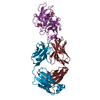


 PDBj
PDBj


 Assembly

 Mass: 18.015 Da / Num. of mol.: 636 / Source method: isolated from a natural source / Formula: H2O
 Sample preparation
 / Beamline: BL7-1
 Processing