 Movie
Movie Controller

[English] 日本語
 Yorodumi
- PDB-1oak: CRYSTAL STRUCTURE OF THE VON WILLEBRAND FACTOR (VWF) A1 DOMAIN IN... -
+ Open data

- Basic information
| Entry | Database: PDB / ID: 1oak | ||||||
|---|---|---|---|---|---|---|---|
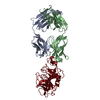
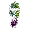
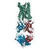
| Title | CRYSTAL STRUCTURE OF THE VON WILLEBRAND FACTOR (VWF) A1 DOMAIN IN COMPLEX WITH THE FUNCTION BLOCKING NMC-4 FAB | ||||||
 Components Components |
| ||||||
 Keywords Keywords | COMPLEX (WILLEBRAND/IMMUNOGLOBULIN) / VON WILLEBRAND FACTOR / GLYCOPROTEIN IBA (A:ALPHA) BINDING / COMPLEX (WILLEBRAND-IMMUNOGLOBULIN) / BLOOD COAGULATION / COMPLEX (WILLEBRAND-IMMUNOGLOBULIN) complex | ||||||
| Function / homology |  Function and homology information Function and homology informationDefective VWF binding to collagen type I / Enhanced cleavage of VWF variant by ADAMTS13 / Defective VWF cleavage by ADAMTS13 variant / Defective F8 binding to von Willebrand factor / Enhanced binding of GP1BA variant to VWF multimer:collagen / Defective binding of VWF variant to GPIb:IX:V / Weibel-Palade body / hemostasis / platelet alpha granule / Platelet Adhesion to exposed collagen ...Defective VWF binding to collagen type I / Enhanced cleavage of VWF variant by ADAMTS13 / Defective VWF cleavage by ADAMTS13 variant / Defective F8 binding to von Willebrand factor / Enhanced binding of GP1BA variant to VWF multimer:collagen / Defective binding of VWF variant to GPIb:IX:V / Weibel-Palade body / hemostasis / platelet alpha granule / Platelet Adhesion to exposed collagen / extracellular matrix structural constituent / GP1b-IX-V activation signalling / p130Cas linkage to MAPK signaling for integrins / Defective F8 cleavage by thrombin / Platelet Aggregation (Plug Formation) / cell-substrate adhesion / GRB2:SOS provides linkage to MAPK signaling for Integrins / positive regulation of intracellular signal transduction / immunoglobulin binding / Integrin cell surface interactions / collagen binding / : / Integrin signaling / platelet alpha granule lumen / Signaling by high-kinase activity BRAF mutants / platelet activation / MAP2K and MAPK activation / response to wounding / integrin binding / blood coagulation / Signaling by RAF1 mutants / Signaling by moderate kinase activity BRAF mutants / Paradoxical activation of RAF signaling by kinase inactive BRAF / Signaling downstream of RAS mutants / Signaling by BRAF and RAF1 fusions / Platelet degranulation / protein-folding chaperone binding / extracellular matrix / protease binding / cell adhesion / endoplasmic reticulum / : / extracellular exosome / extracellular region / identical protein binding Similarity search - Function | ||||||
| Biological species |   Homo sapiens (human) Homo sapiens (human) | ||||||
| Method |  X-RAY DIFFRACTION / SYNCHROTRON / MOLECULAR REPLACEMENT, MIR / Resolution: 2.2 Å X-RAY DIFFRACTION / SYNCHROTRON / MOLECULAR REPLACEMENT, MIR / Resolution: 2.2 Å | ||||||
 Authors Authors | Celikel, R. / Varughese, K.I. | ||||||
 Citation Citation | Journal: Nat.Struct.Biol. / Year: 1998 Title: Crystal structure of the von Willebrand factor A1 domain in complex with the function blocking NMC-4 Fab. Authors: Celikel, R. / Varughese, K.I. / Madhusudan / Yoshioka, A. / Ware, J. / Ruggeri, Z.M. #1: Journal: Blood Cells Mol.Dis. / Year: 1997Title: Crystal Structure of Nmc-4 Fab Anti-Von Willebrand Factor A1 Domain Authors: Celikel, R. / Madhusudan / Varughese, K.I. / Shima, M. / Yoshioka, A. / Ware, J. / Ruggeri, Z.M. | ||||||
| History |
|
- Structure visualization
| Structure viewer | Molecule: MolmilJmol/JSmol |
|---|
- Downloads & links
-Download
| PDBx/mmCIF format | 1oak.cif.gz | 142.3 KB | Display | PDBx/mmCIF format |
|---|---|---|---|---|
| PDB format | pdb1oak.ent.gz | 111.2 KB | Display | PDB format |
| PDBx/mmJSON format | 1oak.json.gz | Tree view | PDBx/mmJSON format | |
| Others |  Other downloads Other downloads |
-Validation report
| Arichive directory | https://data.pdbj.org/pub/pdb/validation_reports/oa/1oakftp://data.pdbj.org/pub/pdb/validation_reports/oa/1oak | HTTPS FTP |
|---|
-Related structure data
| Similar structure data |
|---|

-Links
 PDBj
PDBj


- Assembly
| Deposited unit | 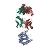
| ||||||||
|---|---|---|---|---|---|---|---|---|---|
| 1 |
| ||||||||
| Unit cell |
|
-Components
| #1: Antibody | Mass: 23461.805 Da / Num. of mol.: 1 Fragment: FAB FRAGMENT, AN ANTI VON WILLEBRAND FACTOR (VWF) A1 DOMAIN Source method: isolated from a genetically manipulated source Details: THE ANTIBODY HEAVY CHAIN CONSISTS OF TWO SEGMENTS, H1 AND H2, BOTH LABELED CHAIN H IN THIS ENTRY Source: (gene. exp.) |
|---|---|
| #2: Antibody | Mass: 23887.551 Da / Num. of mol.: 1 Fragment: FAB FRAGMENT, AN ANTI VON WILLEBRAND FACTOR (VWF) A1 DOMAIN Source method: isolated from a genetically manipulated source Details: THE ANTIBODY HEAVY CHAIN CONSISTS OF TWO SEGMENTS, H1 AND H2, BOTH LABELED CHAIN H IN THIS ENTRY Source: (gene. exp.) |
| #3: Protein | Mass: 22444.135 Da / Num. of mol.: 1 Fragment: A1 DOMAIN RESIDUES 507 - 702, OR GLYCOPROTEIN IBA (A\:ALPHA) BINDING DOMAIN Source method: isolated from a genetically manipulated source Source: (gene. exp.) Homo sapiens (human) / Organ: BLOOD / Plasmid: PET8C / Production host:  |
| #4: Water | ChemComp-HOH /  Mass: 18.015 Da / Num. of mol.: 366 / Source method: isolated from a natural source / Formula: H2O Mass: 18.015 Da / Num. of mol.: 366 / Source method: isolated from a natural source / Formula: H2O |
| Has protein modification | Y |
-Experimental details
-Experiment
| Experiment | Method: X-RAY DIFFRACTION / Number of used crystals: 1 |
|---|
- Sample preparation
| Crystal | Density Matthews: 3.1 Å3/Da / Density % sol: 64 % |
|---|---|
| Crystal grow | pH: 8 / Details: pH 8 |
-Data collection
| Diffraction | Mean temperature: 90 K |
|---|---|
| Diffraction source | Source: SYNCHROTRON / Site: SSRL  / Beamline: BL7-1 / Wavelength: 1.08 / Beamline: BL7-1 / Wavelength: 1.08 |
| Detector | Type: MARRESEARCH / Detector: IMAGE PLATE / Date: Jun 1, 1995 / Details: PT COATED SI FLAT MIRROR BENT FOR VERTICAL FOCUS |
| Radiation | Monochromator: BENT CYLINDRICAL GE(111) / Monochromatic (M) / Laue (L): M / Scattering type: x-ray |
| Radiation wavelength | Wavelength: 1.08 Å / Relative weight: 1 |
| Reflection | Resolution: 2.2→50 Å / Num. obs: 44346 / % possible obs: 99 % / Observed criterion σ(I): 0 / Redundancy: 3.4 % / Rsym value: 0.048 / Net I/σ(I): 18.1 |
| Reflection shell | Resolution: 2.2→2.3 Å / Redundancy: 3.4 % / Rmerge(I) obs: 0.184 / Mean I/σ(I) obs: 6.4 / % possible all: 98.5 |
- Processing
| Software |
| ||||||||||||||||||||||||||||||||||||||||||||||||||||||||||||
|---|---|---|---|---|---|---|---|---|---|---|---|---|---|---|---|---|---|---|---|---|---|---|---|---|---|---|---|---|---|---|---|---|---|---|---|---|---|---|---|---|---|---|---|---|---|---|---|---|---|---|---|---|---|---|---|---|---|---|---|---|---|
| Refinement | Method to determine structure: MOLECULAR REPLACEMENT, MIR / Resolution: 2.2→50 Å / σ(F): 2
| ||||||||||||||||||||||||||||||||||||||||||||||||||||||||||||
| Displacement parameters | Biso mean: 37 Å2
| ||||||||||||||||||||||||||||||||||||||||||||||||||||||||||||
| Refinement step | Cycle: LAST / Resolution: 2.2→50 Å
| ||||||||||||||||||||||||||||||||||||||||||||||||||||||||||||
| Refine LS restraints |
|
