 Movie
Movie Controller

[English] 日本語
 Yorodumi
- PDB-3cfb: High-resolution structure of blue fluorescent antibody EP2-19G2 i... -
+ Open data

- Basic information
| Entry | Database: PDB / ID: 3cfb | ||||||
|---|---|---|---|---|---|---|---|
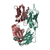
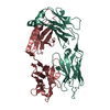
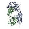
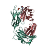
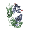
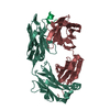
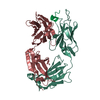
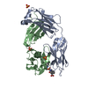
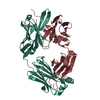
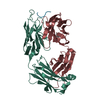
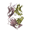
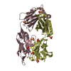
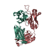
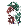
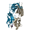
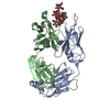
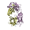
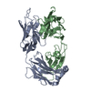
| Title | High-resolution structure of blue fluorescent antibody EP2-19G2 in complex with stilbene hapten at 100K | ||||||
 Components Components |
| ||||||
 Keywords Keywords | IMMUNE SYSTEM / IMMUNOGLOBULIN / BLUE-FLUORESCENT ANTIBODY / HAPTEN COMPLEX / ELECTRON TRANSFER | ||||||
| Function / homology | Immunoglobulins / Immunoglobulin-like / Sandwich / Mainly Beta / 4-(4-STYRYL-PHENYLCARBAMOYL)-BUTYRIC ACID Function and homology information Function and homology information | ||||||
| Biological species |  | ||||||
| Method |  X-RAY DIFFRACTION / SYNCHROTRON / MOLECULAR REPLACEMENT / Resolution: 1.6 Å X-RAY DIFFRACTION / SYNCHROTRON / MOLECULAR REPLACEMENT / Resolution: 1.6 Å | ||||||
 Authors Authors | Debler, E.W. / Wilson, I.A. | ||||||
 Citation Citation | Journal: Science / Year: 2008 Title: Deeply inverted electron-hole recombination in a luminescent antibody-stilbene complex. Authors: Debler, E.W. / Kaufmann, G.F. / Meijler, M.M. / Heine, A. / Mee, J.M. / Pljevaljcic, G. / Di Bilio, A.J. / Schultz, P.G. / Millar, D.P. / Janda, K.D. / Wilson, I.A. / Gray, H.B. / Lerner, R.A. | ||||||
| History |
|
- Structure visualization
| Structure viewer | Molecule: MolmilJmol/JSmol |
|---|
- Downloads & links
-Download
| PDBx/mmCIF format | 3cfb.cif.gz | 194.8 KB | Display | PDBx/mmCIF format |
|---|---|---|---|---|
| PDB format | pdb3cfb.ent.gz | 154.8 KB | Display | PDB format |
| PDBx/mmJSON format | 3cfb.json.gz | Tree view | PDBx/mmJSON format | |
| Others |  Other downloads Other downloads |
-Validation report
| Arichive directory | https://data.pdbj.org/pub/pdb/validation_reports/cf/3cfbftp://data.pdbj.org/pub/pdb/validation_reports/cf/3cfb | HTTPS FTP |
|---|
-Related structure data
| Related structure data | 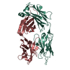 3cfcC  3cfdC 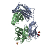 3cfeC 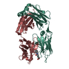 1fl3S S: Starting model for refinement C: citing same article ( |
|---|---|
| Similar structure data |


-Links
 PDBj
PDBj

- Assembly
| Deposited unit | 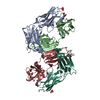
| ||||||||
|---|---|---|---|---|---|---|---|---|---|
| 1 | 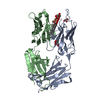
| ||||||||
| 2 | 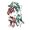
| ||||||||
| Unit cell |
|
-Components
| #1: Antibody | Mass: 24082.723 Da / Num. of mol.: 2 / Fragment: FAB / Source method: isolated from a natural source / Details: PURIFIED FROM ASCITIC FLUID / Source: (natural) Cell: HYBRIDOMA CELLS FROM 129GIX+ SPLEEN CELLS FUSED WITH BALB/C MYELOMA CELLS #2: Antibody | Mass: 22747.699 Da / Num. of mol.: 2 / Fragment: FAB / Source method: isolated from a natural source / Details: PURIFIED FROM ASCITIC FLUID / Source: (natural) Cell: HYBRIDOMA CELLS FROM 129GIX+ SPLEEN CELLS FUSED WITH BALB/C MYELOMA CELLS #3: Chemical |   Mass: 309.359 Da / Num. of mol.: 2 / Source method: obtained synthetically / Formula: C19H19NO3 Mass: 309.359 Da / Num. of mol.: 2 / Source method: obtained synthetically / Formula: C19H19NO3#4: Chemical | ChemComp-GOL /   Mass: 92.094 Da / Num. of mol.: 6 / Source method: obtained synthetically / Formula: C3H8O3 Mass: 92.094 Da / Num. of mol.: 6 / Source method: obtained synthetically / Formula: C3H8O3#5: Water | ChemComp-HOH / |  Mass: 18.015 Da / Num. of mol.: 590 / Source method: isolated from a natural source / Formula: H2O Mass: 18.015 Da / Num. of mol.: 590 / Source method: isolated from a natural source / Formula: H2OHas protein modification | Y | |
|---|
-Experimental details
-Experiment
| Experiment | Method: X-RAY DIFFRACTION / Number of used crystals: 1 |
|---|
- Sample preparation
| Crystal | Density Matthews: 2.61 Å3/Da / Density % sol: 52.93 % |
|---|---|
| Crystal grow | pH: 5.5 Details: 20% PEG 4000, 0.2M KSCN, PH 5.5, VAPOR DIFFUSION, SITTING DROP, TEMPERATURE 298K, pH 5.50 |
-Data collection
| Diffraction | Mean temperature: 100 K |
|---|---|
| Diffraction source | Source: SYNCHROTRON / Site: ALS  / Beamline: 8.2.1 / Wavelength: 0.97622 / Beamline: 8.2.1 / Wavelength: 0.97622 |
| Detector | Type: ADSC QUANTUM 210 / Detector: CCD / Date: Jan 10, 2005 / Details: MONOCHROMATOR |
| Radiation | Monochromator: DOUBLE CRYSTAL, SI(111) / Protocol: SINGLE WAVELENGTH / Monochromatic (M) / Laue (L): M / Scattering type: x-ray |
| Radiation wavelength | Wavelength: 0.97622 Å / Relative weight: 1 |
| Reflection | Resolution: 1.6→50 Å / Num. obs: 121549 / % possible obs: 97.4 % / Observed criterion σ(I): 0 / Redundancy: 3.8 % / Biso Wilson estimate: 58.9 Å2 / Rsym value: 0.052 / Net I/σ(I): 30.3 |
| Reflection shell | Resolution: 1.6→1.66 Å / Redundancy: 3.8 % / Mean I/σ(I) obs: 1.9 / Rsym value: 0.704 / % possible all: 96.2 |
- Processing
| Software |
| |||||||||||||||||||||||||||||||||||||||||||||||||||||||||||||||||||||||||||||||||||||||||||||||||||||||||||||||||||||||||||||||||||||||||||||||||||||||||||||||||||||||||||||||||||||||||||||||||||||||||||||||||||||||||||||||||
|---|---|---|---|---|---|---|---|---|---|---|---|---|---|---|---|---|---|---|---|---|---|---|---|---|---|---|---|---|---|---|---|---|---|---|---|---|---|---|---|---|---|---|---|---|---|---|---|---|---|---|---|---|---|---|---|---|---|---|---|---|---|---|---|---|---|---|---|---|---|---|---|---|---|---|---|---|---|---|---|---|---|---|---|---|---|---|---|---|---|---|---|---|---|---|---|---|---|---|---|---|---|---|---|---|---|---|---|---|---|---|---|---|---|---|---|---|---|---|---|---|---|---|---|---|---|---|---|---|---|---|---|---|---|---|---|---|---|---|---|---|---|---|---|---|---|---|---|---|---|---|---|---|---|---|---|---|---|---|---|---|---|---|---|---|---|---|---|---|---|---|---|---|---|---|---|---|---|---|---|---|---|---|---|---|---|---|---|---|---|---|---|---|---|---|---|---|---|---|---|---|---|---|---|---|---|---|---|---|---|---|---|---|---|---|---|---|---|---|---|---|---|---|---|---|---|---|
| Refinement | Method to determine structure: MOLECULAR REPLACEMENT Starting model: PDB ENTRY 1FL3 Resolution: 1.6→42.03 Å / Cor.coef. Fo:Fc: 0.955 / Cor.coef. Fo:Fc free: 0.931 / SU B: 3.116 / SU ML: 0.059 / TLS residual ADP flag: LIKELY RESIDUAL / Cross valid method: THROUGHOUT / σ(F): 0 / ESU R: 0.088 / ESU R Free: 0.09 / Stereochemistry target values: MAXIMUM LIKELIHOOD / Details: HYDROGENS HAVE BEEN ADDED IN THE RIDING POSITIONS
| |||||||||||||||||||||||||||||||||||||||||||||||||||||||||||||||||||||||||||||||||||||||||||||||||||||||||||||||||||||||||||||||||||||||||||||||||||||||||||||||||||||||||||||||||||||||||||||||||||||||||||||||||||||||||||||||||
| Solvent computation | Ion probe radii: 0.8 Å / Shrinkage radii: 0.8 Å / VDW probe radii: 1.2 Å / Solvent model: BABINET MODEL WITH MASK | |||||||||||||||||||||||||||||||||||||||||||||||||||||||||||||||||||||||||||||||||||||||||||||||||||||||||||||||||||||||||||||||||||||||||||||||||||||||||||||||||||||||||||||||||||||||||||||||||||||||||||||||||||||||||||||||||
| Displacement parameters | Biso mean: 18.11 Å2
| |||||||||||||||||||||||||||||||||||||||||||||||||||||||||||||||||||||||||||||||||||||||||||||||||||||||||||||||||||||||||||||||||||||||||||||||||||||||||||||||||||||||||||||||||||||||||||||||||||||||||||||||||||||||||||||||||
| Refinement step | Cycle: LAST / Resolution: 1.6→42.03 Å
| |||||||||||||||||||||||||||||||||||||||||||||||||||||||||||||||||||||||||||||||||||||||||||||||||||||||||||||||||||||||||||||||||||||||||||||||||||||||||||||||||||||||||||||||||||||||||||||||||||||||||||||||||||||||||||||||||
| Refine LS restraints |
| |||||||||||||||||||||||||||||||||||||||||||||||||||||||||||||||||||||||||||||||||||||||||||||||||||||||||||||||||||||||||||||||||||||||||||||||||||||||||||||||||||||||||||||||||||||||||||||||||||||||||||||||||||||||||||||||||
| LS refinement shell | Resolution: 1.6→1.64 Å / Total num. of bins used: 20
| |||||||||||||||||||||||||||||||||||||||||||||||||||||||||||||||||||||||||||||||||||||||||||||||||||||||||||||||||||||||||||||||||||||||||||||||||||||||||||||||||||||||||||||||||||||||||||||||||||||||||||||||||||||||||||||||||
| Refinement TLS params. | Method: refined / Refine-ID: X-RAY DIFFRACTION
| |||||||||||||||||||||||||||||||||||||||||||||||||||||||||||||||||||||||||||||||||||||||||||||||||||||||||||||||||||||||||||||||||||||||||||||||||||||||||||||||||||||||||||||||||||||||||||||||||||||||||||||||||||||||||||||||||
| Refinement TLS group |
|
