 Movie
Movie Controller

[English] 日本語
 Yorodumi
- PDB-1f58: IGG1 FAB FRAGMENT (58.2) COMPLEX WITH 24-RESIDUE PEPTIDE (RESIDUE... -
+ Open data

- Basic information
| Entry | Database: PDB / ID: 1f58 | ||||||
|---|---|---|---|---|---|---|---|
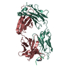
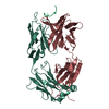
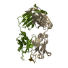
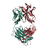
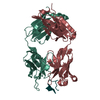
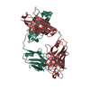
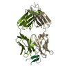
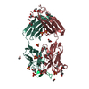
| Title | IGG1 FAB FRAGMENT (58.2) COMPLEX WITH 24-RESIDUE PEPTIDE (RESIDUES 308-333 OF HIV-1 GP120 (MN ISOLATE) WITH ALA TO AIB SUBSTITUTION AT POSITION 323 | ||||||
 Components Components |
| ||||||
 Keywords Keywords | Viral protein/Immune system / IMMUNOGLOBULIN / FAB / HIV-1 / GP120 / V3 / Viral protein-Immune system COMPLEX | ||||||
| Function / homology |  Function and homology information Function and homology informationDectin-2 family / immunoglobulin complex / symbiont-mediated perturbation of host defense response / positive regulation of plasma membrane raft polarization / positive regulation of receptor clustering / host cell endosome membrane / clathrin-dependent endocytosis of virus by host cell / adaptive immune response / immune response / viral protein processing ...Dectin-2 family / immunoglobulin complex / symbiont-mediated perturbation of host defense response / positive regulation of plasma membrane raft polarization / positive regulation of receptor clustering / host cell endosome membrane / clathrin-dependent endocytosis of virus by host cell / adaptive immune response / immune response / viral protein processing / fusion of virus membrane with host plasma membrane / fusion of virus membrane with host endosome membrane / viral envelope / virion attachment to host cell / host cell plasma membrane / virion membrane / structural molecule activity / extracellular region / membrane / metal ion binding / identical protein binding / plasma membrane Similarity search - Function | ||||||
| Biological species |   Human immunodeficiency virus type 1 group M subtype B Human immunodeficiency virus type 1 group M subtype B | ||||||
| Method |  X-RAY DIFFRACTION / MOLECULAR REPLACEMENT / Resolution: 2 Å X-RAY DIFFRACTION / MOLECULAR REPLACEMENT / Resolution: 2 Å | ||||||
 Authors Authors | Stanfield, R.L. / Cabezas, E. / Satterthwait, A.C. / Stura, E.A. / Profy, A.T. / Wilson, I.A. | ||||||
 Citation Citation | Journal: Structure Fold.Des. / Year: 1999 Title: Dual conformations for the HIV-1 gp120 V3 loop in complexes with different neutralizing fabs. Authors: Stanfield, R. / Cabezas, E. / Satterthwait, A. / Stura, E. / Profy, A. / Wilson, I. | ||||||
| History |
|
- Structure visualization
| Structure viewer | Molecule: MolmilJmol/JSmol |
|---|
- Downloads & links
-Download
| PDBx/mmCIF format | 1f58.cif.gz | 102.3 KB | Display | PDBx/mmCIF format |
|---|---|---|---|---|
| PDB format | pdb1f58.ent.gz | 77.9 KB | Display | PDB format |
| PDBx/mmJSON format | 1f58.json.gz | Tree view | PDBx/mmJSON format | |
| Others |  Other downloads Other downloads |
-Validation report
| Arichive directory | https://data.pdbj.org/pub/pdb/validation_reports/f5/1f58ftp://data.pdbj.org/pub/pdb/validation_reports/f5/1f58 | HTTPS FTP |
|---|
-Related structure data


-Links
 PDBj
PDBj


- Assembly
| Deposited unit | 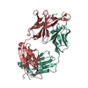
| ||||||||
|---|---|---|---|---|---|---|---|---|---|
| 1 |
| ||||||||
| Unit cell |
|
-Components
| #1: Antibody | Mass: 23573.012 Da / Num. of mol.: 1 / Source method: isolated from a natural source / Source: (natural) |
|---|---|
| #2: Antibody | Mass: 24864.744 Da / Num. of mol.: 1 / Source method: isolated from a natural source / Source: (natural) |
| #3: Protein/peptide | Mass: 2725.223 Da / Num. of mol.: 1 / Fragment: RESIDUES 308-332 FROM HIV-1 GP120 / Source method: obtained synthetically / Details: THE MOLECULE WAS CHEMICALLY SYNTHESIZED Source: (synth.) Human immunodeficiency virus type 1 group M subtype BReferences: UniProt: P05877 |
| #4: Water | ChemComp-HOH /  Mass: 18.015 Da / Num. of mol.: 120 / Source method: isolated from a natural source / Formula: H2O Mass: 18.015 Da / Num. of mol.: 120 / Source method: isolated from a natural source / Formula: H2O |
| Sequence details | THE FAB FRAGMENT IS NUMBERED BY THE CONVENTION OF E. KABAT (E.A. KABAT, T.T. WU, M. REID-MILLER, H. ...THE FAB FRAGMENT IS NUMBERED BY THE CONVENTION |
-Experimental details
-Experiment
| Experiment | Method: X-RAY DIFFRACTION / Number of used crystals: 3 |
|---|
- Sample preparation
| Crystal | Density Matthews: 2.27 Å3/Da / Density % sol: 46 % | ||||||||||||||||||||
|---|---|---|---|---|---|---|---|---|---|---|---|---|---|---|---|---|---|---|---|---|---|
| Crystal grow | pH: 6.3 / Details: pH 6.3 | ||||||||||||||||||||
| Crystal | *PLUS | ||||||||||||||||||||
| Crystal grow | *PLUS Temperature: 22.5 ℃ / Method: vapor diffusion, hanging drop | ||||||||||||||||||||
| Components of the solutions | *PLUS
|
-Data collection
| Diffraction | Mean temperature: 295 K |
|---|---|
| Diffraction source | Source: ROTATING ANODE / Type: RIGAKU RU300 / Wavelength: 1.5418 |
| Detector | Type: RIGAKU RAXIS II / Detector: IMAGE PLATE / Date: Nov 15, 1994 / Details: SUPER LONG MIRRORS (SIEMENS) |
| Radiation | Monochromator: GRAPHITE(RIGAKU), SUPPER LONG MIRRORS (SIEMENS) Protocol: SINGLE WAVELENGTH / Monochromatic (M) / Laue (L): M / Scattering type: x-ray |
| Radiation wavelength | Wavelength: 1.5418 Å / Relative weight: 1 |
| Reflection | Resolution: 2→43.6 Å / Num. obs: 30460 / % possible obs: 99.6 % / Redundancy: 10.4 % / Biso Wilson estimate: 13.6 Å2 / Rmerge(I) obs: 0.106 / Net I/σ(I): 29.5 |
| Reflection shell | Resolution: 2→2.05 Å / Rmerge(I) obs: 0.547 / Mean I/σ(I) obs: 3.4 / % possible all: 98.5 |
| Reflection | *PLUS Num. measured all: 183103 |
| Reflection shell | *PLUS % possible obs: 98.5 % / Num. unique obs: 2014 / Num. measured obs: 9505 |
- Processing
| Software |
| ||||||||||||||||||||||||||||||||||||||||||||||||||||||||||||||||||||||||||||||||
|---|---|---|---|---|---|---|---|---|---|---|---|---|---|---|---|---|---|---|---|---|---|---|---|---|---|---|---|---|---|---|---|---|---|---|---|---|---|---|---|---|---|---|---|---|---|---|---|---|---|---|---|---|---|---|---|---|---|---|---|---|---|---|---|---|---|---|---|---|---|---|---|---|---|---|---|---|---|---|---|---|---|
| Refinement | Method to determine structure: MOLECULAR REPLACEMENT Starting model: FAB 58.2 PORTION OF FAB 58.2/SER-LOOP PEPTIDE COMPLEX Resolution: 2→44 Å / Rfactor Rfree error: 0.005 / Data cutoff high absF: 10000000 / Data cutoff low absF: 0 / Isotropic thermal model: RESTRAINED / Cross valid method: THROUGHOUT / σ(F): 0 / Details: BULK SOLVENT MODEL USED
| ||||||||||||||||||||||||||||||||||||||||||||||||||||||||||||||||||||||||||||||||
| Displacement parameters | Biso mean: 23.4 Å2 | ||||||||||||||||||||||||||||||||||||||||||||||||||||||||||||||||||||||||||||||||
| Refine analyze |
| ||||||||||||||||||||||||||||||||||||||||||||||||||||||||||||||||||||||||||||||||
| Refinement step | Cycle: LAST / Resolution: 2→44 Å
| ||||||||||||||||||||||||||||||||||||||||||||||||||||||||||||||||||||||||||||||||
| Refine LS restraints |
| ||||||||||||||||||||||||||||||||||||||||||||||||||||||||||||||||||||||||||||||||
| LS refinement shell | Resolution: 2→2.05 Å / Rfactor Rfree error: 0.027 / Total num. of bins used: 15
| ||||||||||||||||||||||||||||||||||||||||||||||||||||||||||||||||||||||||||||||||
| Xplor file |
| ||||||||||||||||||||||||||||||||||||||||||||||||||||||||||||||||||||||||||||||||
| Software | *PLUS Name: X-PLOR / Version: 3.851 / Classification: refinement | ||||||||||||||||||||||||||||||||||||||||||||||||||||||||||||||||||||||||||||||||
| Refinement | *PLUS σ(F): 0 / % reflection Rfree: 9.9 % | ||||||||||||||||||||||||||||||||||||||||||||||||||||||||||||||||||||||||||||||||
| Solvent computation | *PLUS | ||||||||||||||||||||||||||||||||||||||||||||||||||||||||||||||||||||||||||||||||
| Displacement parameters | *PLUS Biso mean: 23.4 Å2 | ||||||||||||||||||||||||||||||||||||||||||||||||||||||||||||||||||||||||||||||||
| Refine LS restraints | *PLUS
| ||||||||||||||||||||||||||||||||||||||||||||||||||||||||||||||||||||||||||||||||
| LS refinement shell | *PLUS Rfactor Rfree: 0.382 / % reflection Rfree: 10.1 % / Rfactor Rwork: 0.318 / Rfactor obs: 0.318 |
