 Movie
Movie Controller

[English] 日本語
 Yorodumi
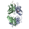
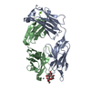
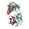
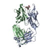
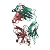
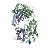
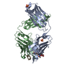
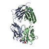
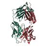
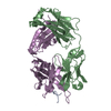
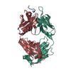
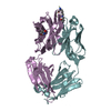
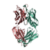
- PDB-5myo: Structure of Pyroglutamate-Abeta-specific Fab c#6 in complex with... -
+ Open data

- Basic information
| Entry | Database: PDB / ID: 5myo | |||||||||
|---|---|---|---|---|---|---|---|---|---|---|
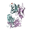
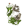
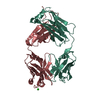
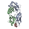
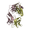
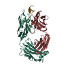
| Title | Structure of Pyroglutamate-Abeta-specific Fab c#6 in complex with human Abeta-pE3-12-PEGb | |||||||||
 Components Components |
| |||||||||
 Keywords Keywords | IMMUNE SYSTEM / Alzheimer's disease / pyroglutamate Abeta / monoclonal antibody / fibrillation | |||||||||
| Function / homology |  Function and homology information Function and homology informationamyloid-beta complex / growth cone lamellipodium / cellular response to norepinephrine stimulus / collateral sprouting in absence of injury / growth cone filopodium / microglia development / hippocampal neuron apoptotic process / Formyl peptide receptors bind formyl peptides and many other ligands / regulation of Wnt signaling pathway / axo-dendritic transport ...amyloid-beta complex / growth cone lamellipodium / cellular response to norepinephrine stimulus / collateral sprouting in absence of injury / growth cone filopodium / microglia development / hippocampal neuron apoptotic process / Formyl peptide receptors bind formyl peptides and many other ligands / regulation of Wnt signaling pathway / axo-dendritic transport / axon midline choice point recognition / regulation of synapse structure or activity / astrocyte activation involved in immune response / NMDA selective glutamate receptor signaling pathway / regulation of spontaneous synaptic transmission / mating behavior / growth factor receptor binding / peptidase activator activity / Insertion of tail-anchored proteins into the endoplasmic reticulum membrane / positive regulation of amyloid fibril formation / Golgi-associated vesicle / PTB domain binding / astrocyte projection / Lysosome Vesicle Biogenesis / Deregulated CDK5 triggers multiple neurodegenerative pathways in Alzheimer's disease models / neuron remodeling / nuclear envelope lumen / dendrite development / regulation of multicellular organism growth / TRAF6 mediated NF-kB activation / positive regulation of protein metabolic process / signaling receptor activator activity / negative regulation of long-term synaptic potentiation / transition metal ion binding / Advanced glycosylation endproduct receptor signaling / The NLRP3 inflammasome / modulation of excitatory postsynaptic potential / main axon / intracellular copper ion homeostasis / ECM proteoglycans / response to insulin-like growth factor stimulus / positive regulation of T cell migration / regulation of presynapse assembly / Notch signaling pathway / neuronal dense core vesicle / swimming behavior / Purinergic signaling in leishmaniasis infection / positive regulation of mitotic cell cycle / adult locomotory behavior / cellular response to manganese ion / positive regulation of chemokine production / extracellular matrix organization / positive regulation of calcium-mediated signaling / axonogenesis / neuron projection maintenance / clathrin-coated pit / Mitochondrial protein degradation / ionotropic glutamate receptor signaling pathway / astrocyte activation / platelet alpha granule lumen / response to interleukin-1 / regulation of neuron apoptotic process / positive regulation of glycolytic process / cellular response to cAMP / cellular response to copper ion / endosome lumen / trans-Golgi network membrane / positive regulation of interleukin-1 beta production / learning / protein serine/threonine kinase binding / dendritic shaft / positive regulation of long-term synaptic potentiation / central nervous system development / Post-translational protein phosphorylation / serine-type endopeptidase inhibitor activity / regulation of long-term neuronal synaptic plasticity / locomotory behavior / microglial cell activation / cellular response to nerve growth factor stimulus / visual learning / positive regulation of non-canonical NF-kappaB signal transduction / TAK1-dependent IKK and NF-kappa-B activation / synapse organization / positive regulation of JNK cascade / recycling endosome / response to lead ion / positive regulation of interleukin-6 production / Golgi lumen / cognition / Regulation of Insulin-like Growth Factor (IGF) transport and uptake by Insulin-like Growth Factor Binding Proteins (IGFBPs) / cellular response to amyloid-beta / neuron projection development / endocytosis / calcium ion transport / positive regulation of inflammatory response / positive regulation of tumor necrosis factor production / Platelet degranulation / heparin binding / regulation of translation / regulation of gene expression Similarity search - Function | |||||||||
| Biological species |   Homo sapiens (human) Homo sapiens (human) | |||||||||
| Method |  X-RAY DIFFRACTION / MOLECULAR REPLACEMENT / molecular replacement / Resolution: 1.59 Å X-RAY DIFFRACTION / MOLECULAR REPLACEMENT / molecular replacement / Resolution: 1.59 Å | |||||||||
 Authors Authors | Parthier, C. / Piechotta, A. / Stubbs, M.T. | |||||||||
 Citation Citation | Journal: J. Biol. Chem. / Year: 2017 Title: Structural and functional analyses of pyroglutamate-amyloid-beta-specific antibodies as a basis for Alzheimer immunotherapy. Authors: Piechotta, A. / Parthier, C. / Kleinschmidt, M. / Gnoth, K. / Pillot, T. / Lues, I. / Demuth, H.U. / Schilling, S. / Rahfeld, J.U. / Stubbs, M.T. | |||||||||
| History |
|
- Structure visualization
| Structure viewer | Molecule: MolmilJmol/JSmol |
|---|
- Downloads & links
-Download
| PDBx/mmCIF format | 5myo.cif.gz | 216.7 KB | Display | PDBx/mmCIF format |
|---|---|---|---|---|
| PDB format | pdb5myo.ent.gz | 169.6 KB | Display | PDB format |
| PDBx/mmJSON format | 5myo.json.gz | Tree view | PDBx/mmJSON format | |
| Others |  Other downloads Other downloads |
-Validation report
| Arichive directory | https://data.pdbj.org/pub/pdb/validation_reports/my/5myoftp://data.pdbj.org/pub/pdb/validation_reports/my/5myo | HTTPS FTP |
|---|
-Related structure data
| Related structure data | 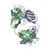 5my4C 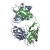 5mykC 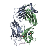 5myxC  1zeaS S: Starting model for refinement C: citing same article ( |
|---|---|
| Similar structure data |

-Links
 PDBj
PDBj


- Assembly
| Deposited unit | 
| ||||||||
|---|---|---|---|---|---|---|---|---|---|
| 1 | 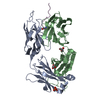
| ||||||||
| 2 | 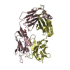
| ||||||||
| Unit cell |
|
-Components
-Protein/peptide , 1 types, 1 molecules E
| #3: Protein/peptide | Mass: 1222.264 Da / Num. of mol.: 1 / Fragment: UNP residues 674-683 / Source method: obtained synthetically / Source: (synth.) Homo sapiens (human) / References: UniProt: P05067 |
|---|
-Antibody , 2 types, 4 molecules ACBD
| #1: Antibody | Mass: 24169.971 Da / Num. of mol.: 2 Source method: isolated from a genetically manipulated source Source: (gene. exp.) #2: Antibody | Mass: 24684.957 Da / Num. of mol.: 2 Source method: isolated from a genetically manipulated source Source: (gene. exp.) |
|---|
-Non-polymers , 3 types, 1161 molecules 


| #4: Chemical | ChemComp-SO4 /  Mass: 96.063 Da / Num. of mol.: 1 / Source method: obtained synthetically / Formula: SO4 Mass: 96.063 Da / Num. of mol.: 1 / Source method: obtained synthetically / Formula: SO4 | ||
|---|---|---|---|
| #5: Chemical |  Mass: 92.094 Da / Num. of mol.: 3 / Source method: obtained synthetically / Formula: C3H8O3 Mass: 92.094 Da / Num. of mol.: 3 / Source method: obtained synthetically / Formula: C3H8O3#6: Water | ChemComp-HOH / | Mass: 18.015 Da / Num. of mol.: 1157 / Source method: isolated from a natural source / Formula: H2O |
-Details
| Has protein modification | Y |
|---|
-Experimental details
-Experiment
| Experiment | Method: X-RAY DIFFRACTION / Number of used crystals: 1 |
|---|
- Sample preparation
| Crystal | Density Matthews: 2.14 Å3/Da / Density % sol: 42.59 % |
|---|---|
| Crystal grow | Temperature: 286 K / Method: vapor diffusion, hanging drop Details: 25.5% w/v PEG 4000 15% glycerol 170 mM ammonium sulfate |
-Data collection
| Diffraction | Mean temperature: 100 K | ||||||||||||||||||||||||||||||||||||||||||||||||||||||||||||||||||||||||||||||||||||||||||||||||||
|---|---|---|---|---|---|---|---|---|---|---|---|---|---|---|---|---|---|---|---|---|---|---|---|---|---|---|---|---|---|---|---|---|---|---|---|---|---|---|---|---|---|---|---|---|---|---|---|---|---|---|---|---|---|---|---|---|---|---|---|---|---|---|---|---|---|---|---|---|---|---|---|---|---|---|---|---|---|---|---|---|---|---|---|---|---|---|---|---|---|---|---|---|---|---|---|---|---|---|---|
| Diffraction source | Source: ROTATING ANODE / Type: RIGAKU MICROMAX-007 / Wavelength: 1.5418 Å | ||||||||||||||||||||||||||||||||||||||||||||||||||||||||||||||||||||||||||||||||||||||||||||||||||
| Detector | Type: RIGAKU SATURN 944+ / Detector: CCD / Date: Sep 8, 2011 | ||||||||||||||||||||||||||||||||||||||||||||||||||||||||||||||||||||||||||||||||||||||||||||||||||
| Radiation | Protocol: SINGLE WAVELENGTH / Monochromatic (M) / Laue (L): M / Scattering type: x-ray | ||||||||||||||||||||||||||||||||||||||||||||||||||||||||||||||||||||||||||||||||||||||||||||||||||
| Radiation wavelength | Wavelength: 1.5418 Å / Relative weight: 1 | ||||||||||||||||||||||||||||||||||||||||||||||||||||||||||||||||||||||||||||||||||||||||||||||||||
| Reflection | Resolution: 1.59→27.189 Å / Num. obs: 99426 / % possible obs: 89.3 % / Observed criterion σ(I): -3 / Redundancy: 2.807 % / Biso Wilson estimate: 14.95 Å2 / Rmerge(I) obs: 0.034 / Rrim(I) all: 0.041 / Χ2: 0.984 / Net I/σ(I): 17.77 | ||||||||||||||||||||||||||||||||||||||||||||||||||||||||||||||||||||||||||||||||||||||||||||||||||
| Reflection shell | Diffraction-ID: 1
|
-Phasing
| Phasing | Method: molecular replacement | |||||||||
|---|---|---|---|---|---|---|---|---|---|---|
| Phasing MR | Rfactor: 56.15 / Model details: Phaser MODE: MR_AUTO
|
- Processing
| Software |
| ||||||||||||||||||||||||||||||||||||||||||||||||||||||||||||||||||||||||||||||||||||||||||||||||||||||||||||||||||||||||||||||||||||||||||||||||||||||||||||||||||||||||||||||||||||||||||
|---|---|---|---|---|---|---|---|---|---|---|---|---|---|---|---|---|---|---|---|---|---|---|---|---|---|---|---|---|---|---|---|---|---|---|---|---|---|---|---|---|---|---|---|---|---|---|---|---|---|---|---|---|---|---|---|---|---|---|---|---|---|---|---|---|---|---|---|---|---|---|---|---|---|---|---|---|---|---|---|---|---|---|---|---|---|---|---|---|---|---|---|---|---|---|---|---|---|---|---|---|---|---|---|---|---|---|---|---|---|---|---|---|---|---|---|---|---|---|---|---|---|---|---|---|---|---|---|---|---|---|---|---|---|---|---|---|---|---|---|---|---|---|---|---|---|---|---|---|---|---|---|---|---|---|---|---|---|---|---|---|---|---|---|---|---|---|---|---|---|---|---|---|---|---|---|---|---|---|---|---|---|---|---|---|---|---|---|
| Refinement | Method to determine structure: MOLECULAR REPLACEMENT Starting model: 1ZEA Resolution: 1.59→27.189 Å / SU ML: 0.18 / Cross valid method: FREE R-VALUE / σ(F): 1.97 / Phase error: 23.76
| ||||||||||||||||||||||||||||||||||||||||||||||||||||||||||||||||||||||||||||||||||||||||||||||||||||||||||||||||||||||||||||||||||||||||||||||||||||||||||||||||||||||||||||||||||||||||||
| Solvent computation | Shrinkage radii: 0.9 Å / VDW probe radii: 1.1 Å | ||||||||||||||||||||||||||||||||||||||||||||||||||||||||||||||||||||||||||||||||||||||||||||||||||||||||||||||||||||||||||||||||||||||||||||||||||||||||||||||||||||||||||||||||||||||||||
| Displacement parameters | Biso max: 63.54 Å2 / Biso mean: 20.2986 Å2 / Biso min: 6.35 Å2 | ||||||||||||||||||||||||||||||||||||||||||||||||||||||||||||||||||||||||||||||||||||||||||||||||||||||||||||||||||||||||||||||||||||||||||||||||||||||||||||||||||||||||||||||||||||||||||
| Refinement step | Cycle: final / Resolution: 1.59→27.189 Å
| ||||||||||||||||||||||||||||||||||||||||||||||||||||||||||||||||||||||||||||||||||||||||||||||||||||||||||||||||||||||||||||||||||||||||||||||||||||||||||||||||||||||||||||||||||||||||||
| Refine LS restraints |
| ||||||||||||||||||||||||||||||||||||||||||||||||||||||||||||||||||||||||||||||||||||||||||||||||||||||||||||||||||||||||||||||||||||||||||||||||||||||||||||||||||||||||||||||||||||||||||
| LS refinement shell | Refine-ID: X-RAY DIFFRACTION / Rfactor Rfree error: 0
|
