 Movie
Movie Controller

[English] 日本語
 Yorodumi
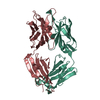
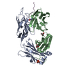
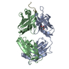
- PDB-4qxu: Novel Inhibition Mechanism of Membrane Metalloprotease by an Exos... -
+ Open data

- Basic information
| Entry | Database: PDB / ID: 4qxu | ||||||
|---|---|---|---|---|---|---|---|
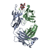
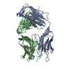
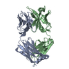
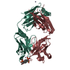
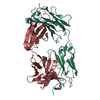
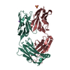
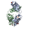
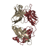
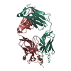
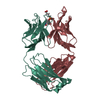
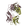
| Title | Novel Inhibition Mechanism of Membrane Metalloprotease by an Exosite-Swiveling Conformational antibody | ||||||
 Components Components |
| ||||||
 Keywords Keywords | IMMUNE SYSTEM / Structural Genomics / PSI-2 / Protein Structure Initiative / Israel Structural Proteomics Center / ISPC / Igg fold | ||||||
| Function / homology |  Function and homology information Function and homology informationmembrane-type matrix metalloproteinase-1 / cellular response to genistein / negative regulation of GDF15-GFRAL signaling pathway / positive regulation of macrophage migration / macropinosome / craniofacial suture morphogenesis / head development / response to odorant / chondrocyte proliferation / positive regulation of protein processing ...membrane-type matrix metalloproteinase-1 / cellular response to genistein / negative regulation of GDF15-GFRAL signaling pathway / positive regulation of macrophage migration / macropinosome / craniofacial suture morphogenesis / head development / response to odorant / chondrocyte proliferation / positive regulation of protein processing / astrocyte cell migration / TGFBR3 PTM regulation / tissue remodeling / negative regulation of focal adhesion assembly / endochondral ossification / embryonic cranial skeleton morphogenesis / intermediate filament cytoskeleton / endothelial cell proliferation / zymogen activation / positive regulation of B cell differentiation / branching morphogenesis of an epithelial tube / endodermal cell differentiation / positive regulation of myotube differentiation / Activation of Matrix Metalloproteinases / metalloaminopeptidase activity / Collagen degradation / collagen catabolic process / negative regulation of Notch signaling pathway / extracellular matrix disassembly / response to mechanical stimulus / regulation of protein localization to plasma membrane / ovarian follicle development / Degradation of the extracellular matrix / extracellular matrix organization / lung development / protein catabolic process / skeletal system development / cell motility / protein processing / metalloendopeptidase activity / male gonad development / response to estrogen / Golgi lumen / integrin binding / melanosome / extracellular matrix / positive regulation of cell growth / angiogenesis / response to oxidative stress / cytoplasmic vesicle / endopeptidase activity / High laminar flow shear stress activates signaling by PIEZO1 and PECAM1:CDH5:KDR in endothelial cells / response to hypoxia / positive regulation of cell migration / serine-type endopeptidase activity / focal adhesion / proteolysis / : / zinc ion binding / nucleus / plasma membrane / cytosol Similarity search - Function | ||||||
| Biological species |   Homo sapiens (human) Homo sapiens (human) | ||||||
| Method |  X-RAY DIFFRACTION / SYNCHROTRON / MOLECULAR REPLACEMENT / Resolution: 2.3 Å X-RAY DIFFRACTION / SYNCHROTRON / MOLECULAR REPLACEMENT / Resolution: 2.3 Å | ||||||
 Authors Authors | Udi, Y. / Grossman, M. / Solomonov, I. / Dym, O. / Rozenberg, H. / Moreno, v. / Cuiniasse, P. / Dive, V. / Arroyo, A.G. / Sagi, I. / Israel Structural Proteomics Center (ISPC) | ||||||
 Citation Citation | Journal: Structure / Year: 2015 Title: Inhibition mechanism of membrane metalloprotease by an exosite-swiveling conformational antibody. Authors: Udi, Y. / Grossman, M. / Solomonov, I. / Dym, O. / Rozenberg, H. / Moreno, V. / Cuniasse, P. / Dive, V. / Arroyo, A.G. / Sagi, I. | ||||||
| History |
|
- Structure visualization
| Structure viewer | Molecule: MolmilJmol/JSmol |
|---|
- Downloads & links
-Download
| PDBx/mmCIF format | 4qxu.cif.gz | 98.8 KB | Display | PDBx/mmCIF format |
|---|---|---|---|---|
| PDB format | pdb4qxu.ent.gz | 75 KB | Display | PDB format |
| PDBx/mmJSON format | 4qxu.json.gz | Tree view | PDBx/mmJSON format | |
| Others |  Other downloads Other downloads |
-Validation report
| Arichive directory | https://data.pdbj.org/pub/pdb/validation_reports/qx/4qxuftp://data.pdbj.org/pub/pdb/validation_reports/qx/4qxu | HTTPS FTP |
|---|
-Related structure data
| Related structure data |  4ouuSC 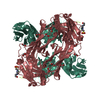 4p3cC  4p3dC S: Starting model for refinement C: citing same article ( |
|---|---|
| Similar structure data |


-Links
 PDBj
PDBj


- Assembly
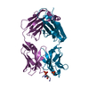
| Deposited unit | 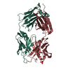
| ||||||||
|---|---|---|---|---|---|---|---|---|---|
| 1 |
| ||||||||
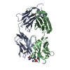
| Unit cell |
|
-Components
| #1: Antibody | Mass: 24124.723 Da / Num. of mol.: 1 Source method: isolated from a genetically manipulated source Source: (gene. exp.)  | ||||
|---|---|---|---|---|---|
| #2: Antibody | Mass: 24995.102 Da / Num. of mol.: 1 Source method: isolated from a genetically manipulated source Source: (gene. exp.) | ||||
| #3: Protein/peptide | Mass: 1330.422 Da / Num. of mol.: 1 / Fragment: peptide (unp residues 218-228) Source method: isolated from a genetically manipulated source Source: (gene. exp.) Homo sapiens (human) / Gene: MMP14 / Production host: References: UniProt: P50281, membrane-type matrix metalloproteinase-1 | ||||
| #4: Chemical |   Mass: 96.063 Da / Num. of mol.: 2 / Source method: obtained synthetically / Formula: SO4 Mass: 96.063 Da / Num. of mol.: 2 / Source method: obtained synthetically / Formula: SO4#5: Water | ChemComp-HOH / |  Mass: 18.015 Da / Num. of mol.: 15 / Source method: isolated from a natural source / Formula: H2O Mass: 18.015 Da / Num. of mol.: 15 / Source method: isolated from a natural source / Formula: H2OHas protein modification | Y | |
-Experimental details
-Experiment
| Experiment | Method: X-RAY DIFFRACTION / Number of used crystals: 1 |
|---|
- Sample preparation
| Crystal | Density Matthews: 2.94 Å3/Da / Density % sol: 58.1 % |
|---|---|
| Crystal grow | Temperature: 298 K / Method: vapor diffusion, hanging drop / pH: 6 Details: 0.1M (NH4)2SO4, 0.01M MgCl2 0.05M MES, 16% PEG 8000, VAPOR DIFFUSION, HANGING DROP, temperature 298K |
-Data collection
| Diffraction | Mean temperature: 298 K |
|---|---|
| Diffraction source | Source: SYNCHROTRON / Site: ESRF  / Beamline: ID29 / Wavelength: 0.97624 Å / Beamline: ID29 / Wavelength: 0.97624 Å |
| Detector | Type: DECTRIS PILATUS 6M / Detector: PIXEL / Date: Mar 18, 2014 |
| Radiation | Protocol: SINGLE WAVELENGTH / Monochromatic (M) / Laue (L): M / Scattering type: x-ray |
| Radiation wavelength | Wavelength: 0.97624 Å / Relative weight: 1 |
| Reflection | Resolution: 2.3→50 Å / Num. all: 27071 / Num. obs: 24287 / % possible obs: 90 % / Observed criterion σ(F): 0 / Observed criterion σ(I): 0 |
| Reflection shell | Resolution: 2.3→2.42 Å / % possible all: 78.9 |
- Processing
| Software |
| ||||||||||||||||||||||||||||||||||||||||||||||||||||||||||||
|---|---|---|---|---|---|---|---|---|---|---|---|---|---|---|---|---|---|---|---|---|---|---|---|---|---|---|---|---|---|---|---|---|---|---|---|---|---|---|---|---|---|---|---|---|---|---|---|---|---|---|---|---|---|---|---|---|---|---|---|---|---|
| Refinement | Method to determine structure: MOLECULAR REPLACEMENT Starting model: pdb entry 4OUU Resolution: 2.3→48.26 Å / Cor.coef. Fo:Fc: 0.913 / Cor.coef. Fo:Fc free: 0.887 / SU B: 8.13 / SU ML: 0.194 / Cross valid method: THROUGHOUT / σ(F): 0 / ESU R: 0.365 / ESU R Free: 0.277 / Stereochemistry target values: MAXIMUM LIKELIHOOD / Details: HYDROGENS HAVE BEEN ADDED IN THE RIDING POSITIONS
| ||||||||||||||||||||||||||||||||||||||||||||||||||||||||||||
| Solvent computation | Ion probe radii: 0.8 Å / Shrinkage radii: 0.8 Å / VDW probe radii: 1.2 Å / Solvent model: MASK | ||||||||||||||||||||||||||||||||||||||||||||||||||||||||||||
| Displacement parameters | Biso mean: 33.424 Å2
| ||||||||||||||||||||||||||||||||||||||||||||||||||||||||||||
| Refinement step | Cycle: LAST / Resolution: 2.3→48.26 Å
| ||||||||||||||||||||||||||||||||||||||||||||||||||||||||||||
| Refine LS restraints |
| ||||||||||||||||||||||||||||||||||||||||||||||||||||||||||||
| LS refinement shell | Resolution: 2.3→2.36 Å / Total num. of bins used: 20
|
