[English] 日本語
Yorodumi
- PDB-6qu9: Fab fragment of an antibody that inhibits polymerisation of alpha... -

+
Open data


ID or keywords:

Loading...

-
Basic information

Entry
Database: PDB / ID: 6qu9
TitleFab fragment of an antibody that inhibits polymerisation of alpha-1-antitrypsin
Components
  • FAB 4B12 heavy chain
  • FAB 4B12 light chain
KeywordsPROTEIN BINDING / Alpha-1 antitrypsin / Z variant / polymers / protein aggregation / monoclonal antibody / Fab fragment / COPD / protease inhibitor / glycoprotein / deficiency
Function / homologyImmunoglobulins / Immunoglobulin-like / Sandwich / Mainly Beta
Function and homology information
Biological speciesMus musculus (house mouse)
MethodX-RAY DIFFRACTION / SYNCHROTRON / MOLECULAR REPLACEMENT / molecular replacement / Resolution: 1.9 Å
AuthorsJagger, A.M. / Heyer-Chauhan, N. / Lomas, D.A. / Irving, J.A.
Funding support United Kingdom, United States, 3items
OrganizationGrant numberCountry
Medical Research Council (MRC, United Kingdom)MR/N024842/1 United Kingdom
Other governmentUCLH/NIHR Biomedical Research Centre United Kingdom
Other privateAlpha-1 Foundation project grant to J. Irving United States
Citation
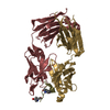
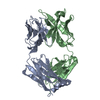
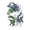
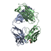
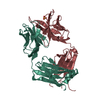
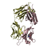
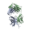
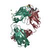
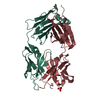
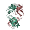
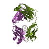
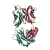
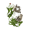
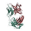
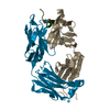
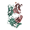
Journal: Sci Adv / Year: 2020
Title: The structural basis for Z α-antitrypsin polymerization in the liver.
Authors: Sarah V Faull / Emma L K Elliston / Bibek Gooptu / Alistair M Jagger / Ibrahim Aldobiyan / Adam Redzej / Magd Badaoui / Nina Heyer-Chauhan / S Tamir Rashid / Gary M Reynolds / David H Adams ...Authors: Sarah V Faull / Emma L K Elliston / Bibek Gooptu / Alistair M Jagger / Ibrahim Aldobiyan / Adam Redzej / Magd Badaoui / Nina Heyer-Chauhan / S Tamir Rashid / Gary M Reynolds / David H Adams / Elena Miranda / Elena V Orlova / James A Irving / David A Lomas /
Abstract: The serpinopathies are among a diverse set of conformational diseases that involve the aberrant self-association of proteins into ordered aggregates. α-Antitrypsin deficiency is the archetypal ...The serpinopathies are among a diverse set of conformational diseases that involve the aberrant self-association of proteins into ordered aggregates. α-Antitrypsin deficiency is the archetypal serpinopathy and results from the formation and deposition of mutant forms of α-antitrypsin as "polymer" chains in liver tissue. No detailed structural analysis has been performed of this material. Moreover, there is little information on the relevance of well-studied artificially induced polymers to these disease-associated molecules. We have isolated polymers from the liver tissue of Z α-antitrypsin homozygotes (E342K) who have undergone transplantation, labeled them using a Fab fragment, and performed single-particle analysis of negative-stain electron micrographs. The data show structural equivalence between heat-induced and ex vivo polymers and that the intersubunit linkage is best explained by a carboxyl-terminal domain swap between molecules of α-antitrypsin.
History
DepositionFeb 26, 2019Deposition site: PDBE / Processing site: PDBE
Revision 1.0Mar 18, 2020Provider: repository / Type: Initial release
Revision 1.1Nov 4, 2020Group: Database references / Category: citation / citation_author
Item: _citation.country / _citation.journal_abbrev ..._citation.country / _citation.journal_abbrev / _citation.journal_id_CSD / _citation.journal_id_ISSN / _citation.journal_volume / _citation.pdbx_database_id_DOI / _citation.pdbx_database_id_PubMed / _citation.title / _citation.year

-
Structure visualization

Structure viewerMolecule:
MolmilJmol/JSmol

Downloads & links

-
Assembly

Deposited unit
H: FAB 4B12 heavy chain
L: FAB 4B12 light chain
A: FAB 4B12 heavy chain
B: FAB 4B12 light chain
hetero molecules


Theoretical massNumber of molelcules
Total (without water)94,1169
Polymers93,7164
Non-polymers3995
Water7,656425
1
H: FAB 4B12 heavy chain
L: FAB 4B12 light chain
hetero molecules


Theoretical massNumber of molelcules
Total (without water)46,9774
Polymers46,8582
Non-polymers1192
Water362
TypeNameSymmetry operationNumber
identity operation1_555x,y,z1
Buried area4000 Å2
ΔGint-47 kcal/mol
Surface area18960 Å2
MethodPISA
2
A: FAB 4B12 heavy chain
B: FAB 4B12 light chain
hetero molecules


Theoretical massNumber of molelcules
Total (without water)47,1385
Polymers46,8582
Non-polymers2803
Water362
TypeNameSymmetry operationNumber
identity operation1_555x,y,z1
Buried area3530 Å2
ΔGint-38 kcal/mol
Surface area18390 Å2
MethodPISA
Unit cell
Length a, b, c (Å)79.268, 105.102, 105.128
Angle α, β, γ (deg.)90.000, 90.000, 90.000
Int Tables number19
Space group name H-MP212121

-
Components

-
Antibody , 2 types, 4 molecules HALB

#1: Antibody FAB 4B12 heavy chain


Mass: 23374.119 Da / Num. of mol.: 2 / Source method: isolated from a natural source / Source: (natural) Mus musculus (house mouse) / Cell line: Hybridoma / Organ: Spleen / Variant: BALB/c
#2: Antibody FAB 4B12 light chain


Mass: 23484.051 Da / Num. of mol.: 2 / Source method: isolated from a natural source / Source: (natural) Mus musculus (house mouse) / Cell line: Hybridoma / Organ: Spleen / Variant: BALB/c

-
Non-polymers , 4 types, 430 molecules

#3: Chemical ChemComp-SO4 / SULFATE ION / Sulfate


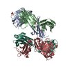
Mass: 96.063 Da / Num. of mol.: 2 / Source method: obtained synthetically / Formula: SO4
#4: Chemical ChemComp-NA / SODIUM ION


Mass: 22.990 Da / Num. of mol.: 1 / Source method: obtained synthetically / Formula: Na
#5: Chemical ChemComp-GOL / GLYCEROL / GLYCERIN / PROPANE-1,2,3-TRIOL / Glycerol


Mass: 92.094 Da / Num. of mol.: 2 / Source method: obtained synthetically / Formula: C3H8O3
#6: Water ChemComp-HOH / water / Water


Mass: 18.015 Da / Num. of mol.: 425 / Source method: isolated from a natural source / Formula: H2O

-
Details

Has ligand of interestN

-
Experimental details

-
Experiment

ExperimentMethod: X-RAY DIFFRACTION / Number of used crystals: 1

-
Sample preparation

CrystalDensity Matthews: 2.35 Å3/Da / Density % sol: 47.65 % / Description: Plate
Crystal growTemperature: 293 K / Method: vapor diffusion, hanging drop / pH: 5.5
Details: 0.1M Bis-Tris pH 5.5, 25% PEG 3350, 0.2M ammonium sulfate

-
Data collection

DiffractionMean temperature: 100 K / Ambient temp details: Cryostream / Serial crystal experiment: N
Diffraction sourceSource: SYNCHROTRON / Site: ESRF / Beamline: ID30B / Wavelength: 0.9763 Å
DetectorType: DECTRIS PILATUS3 6M / Detector: PIXEL / Date: Apr 21, 2018 / Details: Vertical CRL / horizontal eliptical mirror
RadiationMonochromator: Si(111) / Protocol: SINGLE WAVELENGTH / Monochromatic (M) / Laue (L): M / Scattering type: x-ray
Radiation wavelengthWavelength: 0.9763 Å / Relative weight: 1
ReflectionResolution: 1.9→105.1 Å / Num. obs: 69862 / % possible obs: 99.9 % / Redundancy: 6.3 % / CC1/2: 0.997 / Rmerge(I) obs: 0.082 / Rpim(I) all: 0.036 / Rrim(I) all: 0.09 / Χ2: 0.83 / Net I/av σ(I): 9.8 / Net I/σ(I): 9.8
Reflection shell

Diffraction-ID: 1

Resolution (Å)Redundancy (%)Rmerge(I) obsMean I/σ(I) obsNum. unique obsCC1/2Rpim(I) allRrim(I) allΧ2% possible all
1.9-1.946.50.8451.644690.7920.3590.9190.5799.9
9.11-105.15.20.05220.97460.9960.0250.0580.6999.5

-
Phasing

PhasingMethod: molecular replacement
Phasing MR
Highest resolutionLowest resolution
Rotation3.74 Å29.5 Å
Translation3.74 Å29.5 Å

-
Processing

Software
NameVersionClassification
PHENIX1.14-3260refinement
XDS20180126data reduction
Aimless0.6.2data scaling
PHASER2.8.1phasing
PDB_EXTRACT3.24data extraction
RefinementMethod to determine structure: MOLECULAR REPLACEMENT
Starting model: Model generated using RosettaAntibody protocol

Resolution: 1.9→63.287 Å / SU ML: 0.25 / Cross valid method: THROUGHOUT / σ(F): 1.34 / Phase error: 25.94
RfactorNum. reflection% reflection
Rfree0.2328 3388 4.86 %
Rwork0.2019 --
obs0.2034 69773 99.9 %
Solvent computationShrinkage radii: 1 Å / VDW probe radii: 1.2 Å
Displacement parametersBiso max: 100.66 Å2 / Biso mean: 48.0283 Å2 / Biso min: 26.16 Å2
Refinement stepCycle: final / Resolution: 1.9→63.287 Å
ProteinNucleic acidLigandSolventTotal
Num. atoms6378 0 23 425 6826
Biso mean--53.15 49.28 -
Num. residues----848
LS refinement shell

Refine-ID: X-RAY DIFFRACTION / Rfactor Rfree error: 0 / % reflection obs: 100 %

Resolution (Å)Rfactor RfreeNum. reflection RfreeRfactor RworkNum. reflection Rwork
1.9-1.92710.31671460.29992716
1.9271-1.95590.32151300.28742779
1.9559-1.98650.33551390.27772721
1.9865-2.0190.32921560.27222685
2.019-2.05390.32551540.26282738
2.0539-2.09120.28451190.26712769
2.0912-2.13140.31971140.25212758
2.1314-2.17490.24771220.24812719
2.1749-2.22220.28791300.242772
2.2222-2.27390.27521410.23632746
2.2739-2.33080.27991430.23952741
2.3308-2.39380.27581510.23932771
2.3938-2.46430.25331440.24062705
2.4643-2.54380.2791460.23722750
2.5438-2.63470.23541530.23442786
2.6347-2.74020.26351460.23572728
2.7402-2.86490.28311210.23512779
2.8649-3.0160.27731620.22842768
3.016-3.20490.28121410.21022753
3.2049-3.45240.22281540.2092792
3.4524-3.79980.24251640.18522756
3.7998-4.34950.18441070.16782857
4.3495-5.47940.15961530.14822841
5.4794-63.2870.19721520.17822955

+
About Yorodumi

-
News

-
Feb 9, 2022. New format data for meta-information of EMDB entries

New format data for meta-information of EMDB entries

  • Version 3 of the EMDB header file is now the official format.
  • The previous official version 1.9 will be removed from the archive.

Related info.:EMDB header

External links:wwPDB to switch to version 3 of the EMDB data model

-
Aug 12, 2020. Covid-19 info

Covid-19 info

URL: https://pdbj.org/emnavi/covid19.php

New page: Covid-19 featured information page in EM Navigator.

Related info.:Covid-19 info / Mar 5, 2020. Novel coronavirus structure data

+
Mar 5, 2020. Novel coronavirus structure data

Novel coronavirus structure data

Related info.:Yorodumi Speices / Aug 12, 2020. Covid-19 info

External links:COVID-19 featured content - PDBj / Molecule of the Month (242):Coronavirus Proteases

+
Jan 31, 2019. EMDB accession codes are about to change! (news from PDBe EMDB page)

EMDB accession codes are about to change! (news from PDBe EMDB page)

  • The allocation of 4 digits for EMDB accession codes will soon come to an end. Whilst these codes will remain in use, new EMDB accession codes will include an additional digit and will expand incrementally as the available range of codes is exhausted. The current 4-digit format prefixed with “EMD-” (i.e. EMD-XXXX) will advance to a 5-digit format (i.e. EMD-XXXXX), and so on. It is currently estimated that the 4-digit codes will be depleted around Spring 2019, at which point the 5-digit format will come into force.
  • The EM Navigator/Yorodumi systems omit the EMD- prefix.

Related info.:Q: What is EMD? / ID/Accession-code notation in Yorodumi/EM Navigator

External links:EMDB Accession Codes are Changing Soon! / Contact to PDBj

+
Jul 12, 2017. Major update of PDB

Major update of PDB

  • wwPDB released updated PDB data conforming to the new PDBx/mmCIF dictionary.
  • This is a major update changing the version number from 4 to 5, and with Remediation, in which all the entries are updated.
  • In this update, many items about electron microscopy experimental information are reorganized (e.g. em_software).
  • Now, EM Navigator and Yorodumi are based on the updated data.

External links:wwPDB Remediation / Enriched Model Files Conforming to OneDep Data Standards Now Available in the PDB FTP Archive

-
Yorodumi

Thousand views of thousand structures

  • Yorodumi is a browser for structure data from EMDB, PDB, SASBDB, etc.
  • This page is also the successor to EM Navigator detail page, and also detail information page/front-end page for Omokage search.
  • The word "yorodu" (or yorozu) is an old Japanese word meaning "ten thousand". "mi" (miru) is to see.

Related info.:EMDB / PDB / SASBDB / Comparison of 3 databanks / Yorodumi Search / Aug 31, 2016. New EM Navigator & Yorodumi / Yorodumi Papers / Jmol/JSmol / Function and homology information / Changes in new EM Navigator and Yorodumi

Read more