 Movie
Movie Controller

[English] 日本語
 Yorodumi
- PDB-3pp3: Epitope characterization and crystal structure of GA101 provide i... -
+ Open data

- Basic information
| Entry | Database: PDB / ID: 3pp3 | ||||||
|---|---|---|---|---|---|---|---|
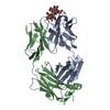
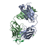
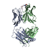
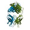
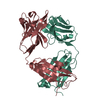
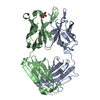
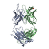
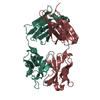
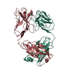
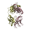
| Title | Epitope characterization and crystal structure of GA101 provide insights into the molecular basis for the type I / type II distinction of anti- CD20 antibodies | ||||||
 Components Components |
| ||||||
 Keywords Keywords | IMMUNE SYSTEM / antibody Fab-fragment Ig-domain / antibody / CD20 | ||||||
| Function / homology | Immunoglobulins / Immunoglobulin-like / Sandwich / Mainly Beta Function and homology information Function and homology information | ||||||
| Biological species |  | ||||||
| Method |  X-RAY DIFFRACTION / SYNCHROTRON / MOLECULAR REPLACEMENT / Resolution: 2.508 Å X-RAY DIFFRACTION / SYNCHROTRON / MOLECULAR REPLACEMENT / Resolution: 2.508 Å | ||||||
 Authors Authors | Hopfner, K.-P. / Lammens, A. | ||||||
 Citation Citation | Journal: Blood / Year: 2011 Title: Epitope characterization and crystal structure of GA101 provide insights into the molecular basis for type I/II distinction of CD20 antibodies. Authors: Niederfellner, G. / Lammens, A. / Mundigl, O. / Georges, G.J. / Schaefer, W. / Schwaiger, M. / Franke, A. / Wiechmann, K. / Jenewein, S. / Slootstra, J.W. / Timmerman, P. / Brannstrom, A. / ...Authors: Niederfellner, G. / Lammens, A. / Mundigl, O. / Georges, G.J. / Schaefer, W. / Schwaiger, M. / Franke, A. / Wiechmann, K. / Jenewein, S. / Slootstra, J.W. / Timmerman, P. / Brannstrom, A. / Lindstrom, F. / Mossner, E. / Umana, P. / Hopfner, K.P. / Klein, C. | ||||||
| History |
|
- Structure visualization
| Structure viewer | Molecule: MolmilJmol/JSmol |
|---|
- Downloads & links
-Download
| PDBx/mmCIF format | 3pp3.cif.gz | 188.7 KB | Display | PDBx/mmCIF format |
|---|---|---|---|---|
| PDB format | pdb3pp3.ent.gz | 148.5 KB | Display | PDB format |
| PDBx/mmJSON format | 3pp3.json.gz | Tree view | PDBx/mmJSON format | |
| Others |  Other downloads Other downloads |
-Validation report
| Arichive directory | https://data.pdbj.org/pub/pdb/validation_reports/pp/3pp3ftp://data.pdbj.org/pub/pdb/validation_reports/pp/3pp3 | HTTPS FTP |
|---|
-Related structure data
| Related structure data |  3pp4C  1t04S C: citing same article ( S: Starting model for refinement |
|---|---|
| Similar structure data |


-Links
 PDBj
PDBj

- Assembly
| Deposited unit | 
| ||||||||
|---|---|---|---|---|---|---|---|---|---|
| 1 | 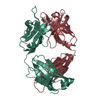
| ||||||||
| 2 | 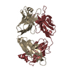
| ||||||||
| Unit cell |
|
-Components
| #1: Antibody | Mass: 23969.881 Da / Num. of mol.: 2 Source method: isolated from a genetically manipulated source Source: (gene. exp.)  Cricetulus griseus (Chinese hamster) Cricetulus griseus (Chinese hamster)#2: Antibody | Mass: 23964.822 Da / Num. of mol.: 2 Source method: isolated from a genetically manipulated source Source: (gene. exp.) Cricetulus griseus (Chinese hamster)#3: Water | ChemComp-HOH / |  Mass: 18.015 Da / Num. of mol.: 527 / Source method: isolated from a natural source / Formula: H2O Mass: 18.015 Da / Num. of mol.: 527 / Source method: isolated from a natural source / Formula: H2OHas protein modification | Y | |
|---|
-Experimental details
-Experiment
| Experiment | Method: X-RAY DIFFRACTION / Number of used crystals: 1 |
|---|
- Sample preparation
| Crystal | Density Matthews: 2.41 Å3/Da / Density % sol: 49.07 % |
|---|---|
| Crystal grow | Temperature: 293 K / Method: vapor diffusion, hanging drop / pH: 7.5 Details: 0.1 M HEPES pH 7.5, 24% (w/v) PEG4000, 0.15 M ammonium sulfate, VAPOR DIFFUSION, HANGING DROP, temperature 293K |
-Data collection
| Diffraction | Mean temperature: 100 K |
|---|---|
| Diffraction source | Source: SYNCHROTRON / Site: ESRF  / Beamline: ID23-1 / Wavelength: 0.9724 Å / Beamline: ID23-1 / Wavelength: 0.9724 Å |
| Detector | Type: MARMOSAIC 225 mm CCD / Detector: CCD / Date: Jun 27, 2008 |
| Radiation | Monochromator: Silicon (1 1 1) channel-cut / Protocol: SINGLE WAVELENGTH / Monochromatic (M) / Laue (L): M / Scattering type: x-ray |
| Radiation wavelength | Wavelength: 0.9724 Å / Relative weight: 1 |
| Reflection | Resolution: 2.5→50 Å / Num. all: 32252 / Num. obs: 31465 / % possible obs: 97.6 % / Observed criterion σ(F): -3 / Redundancy: 3.3 % / Rmerge(I) obs: 0.105 / Rsym value: 0.067 / Net I/σ(I): 12.42 |
| Reflection shell | Resolution: 2.5→2.66 Å / Redundancy: 2.8 % / Rmerge(I) obs: 0.418 / Mean I/σ(I) obs: 3.6 / Rsym value: 0.277 / % possible all: 93.1 |
- Processing
| Software |
| |||||||||||||||||||||||||||||||||||||||||||||||||||||||||||||||||||||||||||||||||||||||||||||||||||||||||
|---|---|---|---|---|---|---|---|---|---|---|---|---|---|---|---|---|---|---|---|---|---|---|---|---|---|---|---|---|---|---|---|---|---|---|---|---|---|---|---|---|---|---|---|---|---|---|---|---|---|---|---|---|---|---|---|---|---|---|---|---|---|---|---|---|---|---|---|---|---|---|---|---|---|---|---|---|---|---|---|---|---|---|---|---|---|---|---|---|---|---|---|---|---|---|---|---|---|---|---|---|---|---|---|---|---|---|
| Refinement | Method to determine structure: MOLECULAR REPLACEMENT Starting model: 1T04 Resolution: 2.508→25 Å / SU ML: 0.4 / σ(F): 1.99 / Stereochemistry target values: ML
| |||||||||||||||||||||||||||||||||||||||||||||||||||||||||||||||||||||||||||||||||||||||||||||||||||||||||
| Solvent computation | Shrinkage radii: 0.72 Å / VDW probe radii: 1 Å / Solvent model: FLAT BULK SOLVENT MODEL / Bsol: 46.581 Å2 / ksol: 0.341 e/Å3 | |||||||||||||||||||||||||||||||||||||||||||||||||||||||||||||||||||||||||||||||||||||||||||||||||||||||||
| Displacement parameters |
| |||||||||||||||||||||||||||||||||||||||||||||||||||||||||||||||||||||||||||||||||||||||||||||||||||||||||
| Refinement step | Cycle: LAST / Resolution: 2.508→25 Å
| |||||||||||||||||||||||||||||||||||||||||||||||||||||||||||||||||||||||||||||||||||||||||||||||||||||||||
| Refine LS restraints |
| |||||||||||||||||||||||||||||||||||||||||||||||||||||||||||||||||||||||||||||||||||||||||||||||||||||||||
| LS refinement shell |
|
