 Movie
Movie Controller

[English] 日本語
 Yorodumi
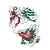
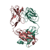
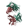
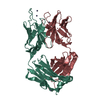
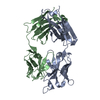
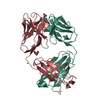
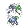
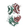
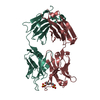
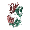
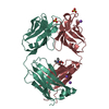
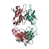
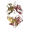
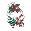
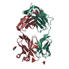
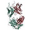
- PDB-1t04: Three dimensional structure of a humanized anti-IFN-Gamma Fab in ... -
+ Open data

- Basic information
| Entry | Database: PDB / ID: 1t04 | |||||||||
|---|---|---|---|---|---|---|---|---|---|---|
| Title | Three dimensional structure of a humanized anti-IFN-Gamma Fab in C2 space group | |||||||||
 Components Components |
| |||||||||
 Keywords Keywords | IMMUNE SYSTEM / Antibody Engineering / Humanized and Chimeric Antibody / FAB / three-dimensional structure / Gamma-interferon | |||||||||
| Function / homology | Immunoglobulins / Immunoglobulin-like / Sandwich / Mainly Beta / :  Function and homology information Function and homology information | |||||||||
| Biological species |  Homo sapiens (human) Homo sapiens (human) | |||||||||
| Method |  X-RAY DIFFRACTION / MOLECULAR REPLACEMENT / Resolution: 3 Å X-RAY DIFFRACTION / MOLECULAR REPLACEMENT / Resolution: 3 Å | |||||||||
 Authors Authors | Bourne, P.C. / Terzyan, S.S. / Cloud, G. / Landolfi, N.F. / Vasquez, M. / Edmundson, A.B. | |||||||||
 Citation Citation | Journal: Acta Crystallogr.,Sect.D / Year: 2004 Title: Three-dimensional structures of a humanized anti-IFN-gamma Fab (HuZAF) in two crystal forms. Authors: Bourne, P.C. / Terzyan, S.S. / Cloud, G. / Landolfi, N.F. / Vasquez, M. / Edmundson, A.B. | |||||||||
| History |
| |||||||||
| Remark 999 | SEQUENCE THERE IS NO SEQUENCE DATABASE REFERENCE FOR THIS ENTRY. THE SEQUENCE WAS DETERMINED IN THE ...SEQUENCE THERE IS NO SEQUENCE DATABASE REFERENCE FOR THIS ENTRY. THE SEQUENCE WAS DETERMINED IN THE PROTEIN DESIGN LABORATORY. |
- Structure visualization
| Structure viewer | Molecule: MolmilJmol/JSmol |
|---|
- Downloads & links
-Download
| PDBx/mmCIF format | 1t04.cif.gz | 172.9 KB | Display | PDBx/mmCIF format |
|---|---|---|---|---|
| PDB format | pdb1t04.ent.gz | 139 KB | Display | PDB format |
| PDBx/mmJSON format | 1t04.json.gz | Tree view | PDBx/mmJSON format | |
| Others |  Other downloads Other downloads |
-Validation report
| Arichive directory | https://data.pdbj.org/pub/pdb/validation_reports/t0/1t04ftp://data.pdbj.org/pub/pdb/validation_reports/t0/1t04 | HTTPS FTP |
|---|
-Related structure data
| Related structure data |  1t3fSC S: Starting model for refinement C: citing same article ( |
|---|---|
| Similar structure data |


-Links
 PDBj
PDBj

- Assembly
| Deposited unit | 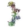
| ||||||||
|---|---|---|---|---|---|---|---|---|---|
| 1 | 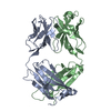
| ||||||||
| 2 | 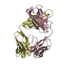
| ||||||||
| Unit cell |
|
-Components
| #1: Antibody | Mass: 23430.879 Da / Num. of mol.: 2 Source method: isolated from a genetically manipulated source Details: The source is engineered human constant and framework regions with CDRs from mouse. Source: (gene. exp.) Homo sapiens (human) / References: UniProt: Q6GMW1#2: Antibody | Mass: 23377.156 Da / Num. of mol.: 2 Source method: isolated from a genetically manipulated source Details: The source is engineered human constant and framework regions with CDRs from mouse. Source: (gene. exp.) Homo sapiens (human)#3: Water | ChemComp-HOH / |  Mass: 18.015 Da / Num. of mol.: 47 / Source method: isolated from a natural source / Formula: H2O Mass: 18.015 Da / Num. of mol.: 47 / Source method: isolated from a natural source / Formula: H2OHas protein modification | Y | |
|---|
-Experimental details
-Experiment
| Experiment | Method: X-RAY DIFFRACTION / Number of used crystals: 1 |
|---|
- Sample preparation
| Crystal | Density Matthews: 2.7 Å3/Da / Density % sol: 46 % |
|---|---|
| Crystal grow | Temperature: 288 K / pH: 5.6 Details: PEG8000, (NH4)2SO4, MgCl2, MES, pH 5.60, VAPOR DIFFUSION, SITTING DROP, temperature 288K |
-Data collection
| Diffraction | Mean temperature: 100 K |
|---|---|
| Diffraction source | Source: ROTATING ANODE / Type: RIGAKU RUH3R / Wavelength: 1.54 |
| Detector | Type: MARRESEARCH / Detector: IMAGE PLATE / Date: Apr 20, 2003 / Details: OSMIC BLUE OPTICS |
| Radiation | Monochromator: OSMIC BLUE OPTICS / Protocol: SINGLE WAVELENGTH / Monochromatic (M) / Laue (L): M / Scattering type: x-ray |
| Radiation wavelength | Wavelength: 1.54 Å / Relative weight: 1 |
| Reflection | Resolution: 3→25 Å / Num. obs: 19324 / % possible obs: 97.1 % / Observed criterion σ(I): -3 / Redundancy: 2.49 % / Biso Wilson estimate: 51.72 Å2 / Rmerge(I) obs: 0.146 / Net I/σ(I): 6.14 |
| Reflection shell | Resolution: 3→3.11 Å / Rmerge(I) obs: 0.387 / Mean I/σ(I) obs: 2.27 / % possible all: 96.9 |
- Processing
| Software |
| ||||||||||||||||||||||||||||||||||||||||||||||||||||||||||||
|---|---|---|---|---|---|---|---|---|---|---|---|---|---|---|---|---|---|---|---|---|---|---|---|---|---|---|---|---|---|---|---|---|---|---|---|---|---|---|---|---|---|---|---|---|---|---|---|---|---|---|---|---|---|---|---|---|---|---|---|---|---|
| Refinement | Method to determine structure: MOLECULAR REPLACEMENT Starting model: PDB ENTRY 1T3F Resolution: 3→25 Å / Isotropic thermal model: OVERALL ANISOTROPIC / Cross valid method: THROUGHOUT / σ(F): 0 / Stereochemistry target values: ENGH & HUBER
| ||||||||||||||||||||||||||||||||||||||||||||||||||||||||||||
| Solvent computation | Solvent model: CNS / Bsol: 31.82 Å2 / ksol: 0.36 e/Å3 | ||||||||||||||||||||||||||||||||||||||||||||||||||||||||||||
| Displacement parameters | Biso mean: 28.27 Å2
| ||||||||||||||||||||||||||||||||||||||||||||||||||||||||||||
| Refinement step | Cycle: LAST / Resolution: 3→25 Å
| ||||||||||||||||||||||||||||||||||||||||||||||||||||||||||||
| Refine LS restraints |
| ||||||||||||||||||||||||||||||||||||||||||||||||||||||||||||
| Xplor file |
|
