 Movie
Movie Controller

[English] 日本語
 Yorodumi
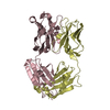
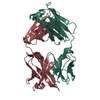
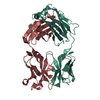
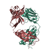
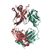
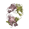
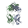
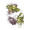
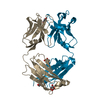
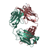
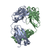
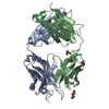
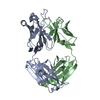
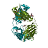
- PDB-1b2w: COMPARISON OF THE THREE-DIMENSIONAL STRUCTURES OF A HUMANIZED AND... -
+ Open data

- Basic information
| Entry | Database: PDB / ID: 1b2w | |||||||||
|---|---|---|---|---|---|---|---|---|---|---|
| Title | COMPARISON OF THE THREE-DIMENSIONAL STRUCTURES OF A HUMANIZED AND A CHIMERIC FAB OF AN ANTI-GAMMA-INTERFERON ANTIBODY | |||||||||
 Components Components |
| |||||||||
 Keywords Keywords | IMMUNE SYSTEM / ANTIBODY ENGINEERING / HUMANIZED AND CHIMERIC ANTIBODY / FAB / THREE-DIMENSIONAL STRYCTURE / GAMMA-INTERFERON | |||||||||
| Function / homology | Immunoglobulins / Immunoglobulin-like / Sandwich / Mainly Beta Function and homology information Function and homology information | |||||||||
| Biological species |   Homo sapiens (human) Homo sapiens (human) | |||||||||
| Method |  X-RAY DIFFRACTION / MOLECULAR REPLACEMENT / Resolution: 2.9 Å X-RAY DIFFRACTION / MOLECULAR REPLACEMENT / Resolution: 2.9 Å | |||||||||
 Authors Authors | Fan, Z. / Shan, L. / Goldsteen, B.Z. / Guddat, L.W. / Thakur, A. / Landolfi, N.F. / Co, M.S. / Vasquez, M. / Queen, C. / Ramsland, P.A. / Edmundson, A.B. | |||||||||
 Citation Citation | Journal: J.Mol.Recog. / Year: 1999 Title: Comparison of the three-dimensional structures of a humanized and a chimeric Fab of an anti-gamma-interferon antibody. Authors: Fan, Z.C. / Shan, L. / Goldsteen, B.Z. / Guddat, L.W. / Thakur, A. / Landolfi, N.F. / Co, M.S. / Vasquez, M. / Queen, C. / Ramsland, P.A. / Edmundson, A.B. | |||||||||
| History |
|
- Structure visualization
| Structure viewer | Molecule: MolmilJmol/JSmol |
|---|
- Downloads & links
-Download
| PDBx/mmCIF format | 1b2w.cif.gz | 86.4 KB | Display | PDBx/mmCIF format |
|---|---|---|---|---|
| PDB format | pdb1b2w.ent.gz | 66.7 KB | Display | PDB format |
| PDBx/mmJSON format | 1b2w.json.gz | Tree view | PDBx/mmJSON format | |
| Others |  Other downloads Other downloads |
-Validation report
| Arichive directory | https://data.pdbj.org/pub/pdb/validation_reports/b2/1b2wftp://data.pdbj.org/pub/pdb/validation_reports/b2/1b2w | HTTPS FTP |
|---|
-Related structure data
| Related structure data | 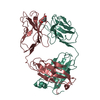 1b4jC 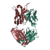 1dfbS S: Starting model for refinement C: citing same article ( |
|---|---|
| Similar structure data |


-Links
 PDBj
PDBj

- Assembly
| Deposited unit | 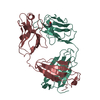
| ||||||||
|---|---|---|---|---|---|---|---|---|---|
| 1 |
| ||||||||
| Unit cell |
|
-Components
| #1: Antibody | Mass: 23430.879 Da / Num. of mol.: 1 / Fragment: V DOMAIN AND C DOMAIN Source method: isolated from a genetically manipulated source Source: (gene. exp.) Mus musculus, Homo sapiens |
|---|---|
| #2: Antibody | Mass: 23800.799 Da / Num. of mol.: 1 / Fragment: V DOMAIN AND C DOMAIN Source method: isolated from a genetically manipulated source Source: (gene. exp.) Mus musculus, Homo sapiens |
| Has protein modification | Y |
-Experimental details
-Experiment
| Experiment | Method: X-RAY DIFFRACTION / Number of used crystals: 1 |
|---|
- Sample preparation
| Crystal | Density Matthews: 2.85 Å3/Da / Density % sol: 57 % | ||||||||||||||||||||||||||||||
|---|---|---|---|---|---|---|---|---|---|---|---|---|---|---|---|---|---|---|---|---|---|---|---|---|---|---|---|---|---|---|---|
| Crystal grow | pH: 6.2 Details: 6.6% PEG 8000(W/V), IN 0.05 M NACL AND 0.05% SODIUM AZIDE., pH 6.2 | ||||||||||||||||||||||||||||||
| Components of the solutions |
| ||||||||||||||||||||||||||||||
| Crystal | *PLUS | ||||||||||||||||||||||||||||||
| Crystal grow | *PLUS Temperature: 18-20 ℃ / Method: vapor diffusion | ||||||||||||||||||||||||||||||
| Components of the solutions | *PLUS
|
-Data collection
| Diffraction | Mean temperature: 286 K |
|---|---|
| Diffraction source | Source: ROTATING ANODE / Type: SIEMENS / Wavelength: 1.5418 |
| Detector | Type: SIEMENS / Detector: AREA DETECTOR / Date: Jul 1, 1994 |
| Radiation | Monochromator: NI FILTER / Protocol: SINGLE WAVELENGTH / Monochromatic (M) / Laue (L): M / Scattering type: x-ray |
| Radiation wavelength | Wavelength: 1.5418 Å / Relative weight: 1 |
| Reflection | Resolution: 2.79→20 Å / Num. obs: 11491 / % possible obs: 81.7 % / Redundancy: 2.2 % / Biso Wilson estimate: 38.8 Å2 / Rmerge(I) obs: 0.093 / Rsym value: 0.093 / Net I/σ(I): 8.3 |
| Reflection shell | Resolution: 2.79→2.96 Å / Redundancy: 1.2 % / Rmerge(I) obs: 0.41 / Mean I/σ(I) obs: 1 / Rsym value: 0.41 / % possible all: 0.28 |
| Reflection | *PLUS % possible obs: 67.2 % |
- Processing
| Software |
| ||||||||||||||||||||||||||||||||||||||||||||||||||||||||||||
|---|---|---|---|---|---|---|---|---|---|---|---|---|---|---|---|---|---|---|---|---|---|---|---|---|---|---|---|---|---|---|---|---|---|---|---|---|---|---|---|---|---|---|---|---|---|---|---|---|---|---|---|---|---|---|---|---|---|---|---|---|---|
| Refinement | Method to determine structure: MOLECULAR REPLACEMENT Starting model: 1DFB Resolution: 2.9→8 Å / Rfactor Rfree error: 0.011 / Data cutoff high absF: 1000000 / Data cutoff low absF: 0.001 / Isotropic thermal model: RESTRAINED / σ(F): 0
| ||||||||||||||||||||||||||||||||||||||||||||||||||||||||||||
| Displacement parameters | Biso mean: 30 Å2 | ||||||||||||||||||||||||||||||||||||||||||||||||||||||||||||
| Refine analyze | Luzzati coordinate error obs: 0.31 Å / Luzzati d res low obs: 8 Å | ||||||||||||||||||||||||||||||||||||||||||||||||||||||||||||
| Refinement step | Cycle: LAST / Resolution: 2.9→8 Å
| ||||||||||||||||||||||||||||||||||||||||||||||||||||||||||||
| Refine LS restraints |
| ||||||||||||||||||||||||||||||||||||||||||||||||||||||||||||
| LS refinement shell | Resolution: 2.9→3.03 Å / Rfactor Rfree error: 0.052 / Total num. of bins used: 8
| ||||||||||||||||||||||||||||||||||||||||||||||||||||||||||||
| Xplor file |
| ||||||||||||||||||||||||||||||||||||||||||||||||||||||||||||
| Software | *PLUS Name: X-PLOR / Version: 3.8 / Classification: refinement | ||||||||||||||||||||||||||||||||||||||||||||||||||||||||||||
| Refinement | *PLUS % reflection Rfree: 10 % / Rfactor obs: 0.188 / Rfactor Rfree: 0.331 | ||||||||||||||||||||||||||||||||||||||||||||||||||||||||||||
| Solvent computation | *PLUS | ||||||||||||||||||||||||||||||||||||||||||||||||||||||||||||
| Displacement parameters | *PLUS Biso mean: 30 Å2 | ||||||||||||||||||||||||||||||||||||||||||||||||||||||||||||
| Refine LS restraints | *PLUS
|
