 Movie
Movie Controller

[English] 日本語
 Yorodumi
- PDB-5it2: Structure of a transglutaminase 2-specific autoantibody 693-10-B0... -
+ Open data

- Basic information
| Entry | Database: PDB / ID: 5it2 | ||||||
|---|---|---|---|---|---|---|---|
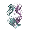
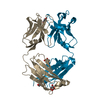
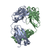
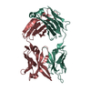
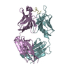
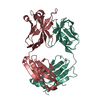
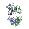
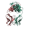
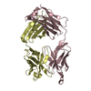
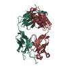
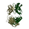
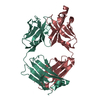
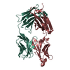
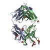
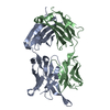
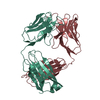
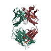
| Title | Structure of a transglutaminase 2-specific autoantibody 693-10-B06 Fab fragment | ||||||
 Components Components |
| ||||||
 Keywords Keywords | IMMUNE SYSTEM / Transglutaminase 2 / autoantibody / Fab fragment | ||||||
| Function / homology | Immunoglobulins / Immunoglobulin-like / Sandwich / Mainly Beta Function and homology information Function and homology information | ||||||
| Biological species |  Homo sapiens (human) Homo sapiens (human) | ||||||
| Method |  X-RAY DIFFRACTION / SYNCHROTRON / MOLECULAR REPLACEMENT / Resolution: 1.7 Å X-RAY DIFFRACTION / SYNCHROTRON / MOLECULAR REPLACEMENT / Resolution: 1.7 Å | ||||||
 Authors Authors | Chen, X. / Dalhus, B. / Hnida, K. / Iversen, R. / Sollid, L.M. | ||||||
 Citation Citation | Journal: Nat. Med. / Year: 2012 Title: High abundance of plasma cells secreting transglutaminase 2-specific IgA autoantibodies with limited somatic hypermutation in celiac disease intestinal lesions Authors: Di Niro, R. / Mesin, L. / Zheng, N.Y. / Stamnaes, J. / Morrissey, M. / Lee, J.H. / Huang, M. / Iversen, R. / du Pre, M.F. / Qiao, S.W. / Lundin, K.E. / Wilson, P.C. / Sollid, L.M. | ||||||
| History |
|
- Structure visualization
| Structure viewer | Molecule: MolmilJmol/JSmol |
|---|
- Downloads & links
-Download
| PDBx/mmCIF format | 5it2.cif.gz | 196.4 KB | Display | PDBx/mmCIF format |
|---|---|---|---|---|
| PDB format | pdb5it2.ent.gz | 157 KB | Display | PDB format |
| PDBx/mmJSON format | 5it2.json.gz | Tree view | PDBx/mmJSON format | |
| Others |  Other downloads Other downloads |
-Validation report
| Arichive directory | https://data.pdbj.org/pub/pdb/validation_reports/it/5it2ftp://data.pdbj.org/pub/pdb/validation_reports/it/5it2 | HTTPS FTP |
|---|
-Related structure data
| Related structure data | 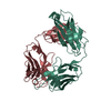 4zd3S S: Starting model for refinement |
|---|---|
| Similar structure data |


-Links
 PDBj
PDBj

- Assembly
| Deposited unit | 
| ||||||||
|---|---|---|---|---|---|---|---|---|---|
| 1 |
| ||||||||
| Unit cell |
|
-Components
| #1: Antibody | Mass: 24363.463 Da / Num. of mol.: 1 Source method: isolated from a genetically manipulated source Source: (gene. exp.) Homo sapiens (human) / Production host: Homo sapiens (human) |
|---|---|
| #2: Antibody | Mass: 23526.066 Da / Num. of mol.: 1 Source method: isolated from a genetically manipulated source Source: (gene. exp.) Homo sapiens (human) / Production host: Homo sapiens (human) |
| #3: Water | ChemComp-HOH /  Mass: 18.015 Da / Num. of mol.: 637 / Source method: isolated from a natural source / Formula: H2O Mass: 18.015 Da / Num. of mol.: 637 / Source method: isolated from a natural source / Formula: H2O |
| Has protein modification | Y |
-Experimental details
-Experiment
| Experiment | Method: X-RAY DIFFRACTION / Number of used crystals: 1 |
|---|
- Sample preparation
| Crystal | Density Matthews: 2.59 Å3/Da / Density % sol: 52.45 % |
|---|---|
| Crystal grow | Temperature: 293 K / Method: vapor diffusion, hanging drop / pH: 8.5 / Details: 0.1M Tris, pH=8.5, 20%v/v Ethanol |
-Data collection
| Diffraction | Mean temperature: 100 K |
|---|---|
| Diffraction source | Source: SYNCHROTRON / Site: ESRF  / Beamline: ID23-2 / Wavelength: 0.8726 Å / Beamline: ID23-2 / Wavelength: 0.8726 Å |
| Detector | Type: DECTRIS PILATUS3 2M / Detector: PIXEL / Date: Feb 20, 2014 |
| Radiation | Protocol: SINGLE WAVELENGTH / Monochromatic (M) / Laue (L): M / Scattering type: x-ray |
| Radiation wavelength | Wavelength: 0.8726 Å / Relative weight: 1 |
| Reflection | Resolution: 1.7→54.24 Å / Num. obs: 54158 / % possible obs: 98.1 % / Redundancy: 3.6 % / Rmerge(I) obs: 0.082 / Rsym value: 0.082 / Net I/σ(I): 25.2 |
| Reflection shell | Resolution: 1.7→1.73 Å / Redundancy: 3.3 % / Rmerge(I) obs: 0.509 / Mean I/σ(I) obs: 2.1 / % possible all: 99.2 |
- Processing
| Software |
| ||||||||||||||||||||
|---|---|---|---|---|---|---|---|---|---|---|---|---|---|---|---|---|---|---|---|---|---|
| Refinement | Method to determine structure: MOLECULAR REPLACEMENT Starting model: 4ZD3 Resolution: 1.7→43.867 Å / Cross valid method: FREE R-VALUE
| ||||||||||||||||||||
| Refinement step | Cycle: LAST / Resolution: 1.7→43.867 Å
| ||||||||||||||||||||
| LS refinement shell | Resolution: 1.7→1.7293 Å / Total num. of bins used: 20
|
