 Movie
Movie Controller


+ Open data

- Basic information
| Entry | Database: PDB / ID: 3eo9 | ||||||
|---|---|---|---|---|---|---|---|
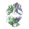
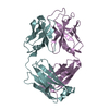
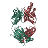
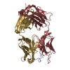
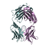
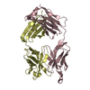
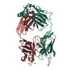
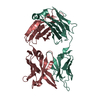
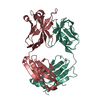
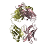
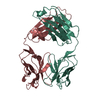
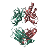
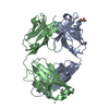
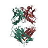
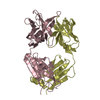
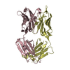
| Title | Crystal structure the Fab fragment of Efalizumab | ||||||
 Components Components |
| ||||||
 Keywords Keywords | IMMUNE SYSTEM / Efalizumab / Fab / antibody | ||||||
| Function / homology | Immunoglobulins / Immunoglobulin-like / Sandwich / Mainly Beta Function and homology information Function and homology information | ||||||
| Biological species |  Homo sapiens (human) Homo sapiens (human) | ||||||
| Method |  X-RAY DIFFRACTION / MOLECULAR REPLACEMENT / Resolution: 1.8 Å X-RAY DIFFRACTION / MOLECULAR REPLACEMENT / Resolution: 1.8 Å | ||||||
 Authors Authors | Li, S. / Ding, J. | ||||||
 Citation Citation | Journal: Proc.Natl.Acad.Sci.USA / Year: 2009 Title: Efalizumab binding to the LFA-1 alphaL I domain blocks ICAM-1 binding via steric hindrance. Authors: Li, S. / Wang, H. / Peng, B. / Zhang, M. / Zhang, D. / Hou, S. / Guo, Y. / Ding, J. | ||||||
| History |
|
- Structure visualization
| Structure viewer | Molecule: MolmilJmol/JSmol |
|---|
- Downloads & links
-Download
| PDBx/mmCIF format | 3eo9.cif.gz | 188.9 KB | Display | PDBx/mmCIF format |
|---|---|---|---|---|
| PDB format | pdb3eo9.ent.gz | 150.6 KB | Display | PDB format |
| PDBx/mmJSON format | 3eo9.json.gz | Tree view | PDBx/mmJSON format | |
| Others |  Other downloads Other downloads |
-Validation report
| Arichive directory | https://data.pdbj.org/pub/pdb/validation_reports/eo/3eo9ftp://data.pdbj.org/pub/pdb/validation_reports/eo/3eo9 | HTTPS FTP |
|---|
-Related structure data
| Related structure data |  3eoaC  3eobC 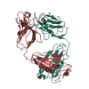 1b2wS C: citing same article ( S: Starting model for refinement |
|---|---|
| Similar structure data |


-Links
 PDBj
PDBj

- Assembly
| Deposited unit | 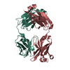
| ||||||||
|---|---|---|---|---|---|---|---|---|---|
| 1 |
| ||||||||
| Unit cell |
|
-Components
| #1: Antibody | Mass: 23436.068 Da / Num. of mol.: 1 Source method: isolated from a genetically manipulated source Source: (gene. exp.) Homo sapiens (human) / Gene: IGG1 |
|---|---|
| #2: Antibody | Mass: 23622.498 Da / Num. of mol.: 1 Source method: isolated from a genetically manipulated source Source: (gene. exp.) Homo sapiens (human) / Gene: IGG1 |
| #3: Water | ChemComp-HOH /  Mass: 18.015 Da / Num. of mol.: 625 / Source method: isolated from a natural source / Formula: H2O Mass: 18.015 Da / Num. of mol.: 625 / Source method: isolated from a natural source / Formula: H2O |
| Has protein modification | Y |
-Experimental details
-Experiment
| Experiment | Method: X-RAY DIFFRACTION / Number of used crystals: 1 |
|---|
- Sample preparation
| Crystal | Density Matthews: 2.73 Å3/Da / Density % sol: 55.01 % |
|---|---|
| Crystal grow | Temperature: 277 K / Method: vapor diffusion, hanging drop / pH: 5 Details: 15mM citric acid, 12% w/v PEG 3350, pH 5.0, VAPOR DIFFUSION, HANGING DROP, temperature 277K |
-Data collection
| Diffraction | Mean temperature: 100 K |
|---|---|
| Diffraction source | Source: ROTATING ANODE / Type: RIGAKU / Wavelength: 1.5418 Å |
| Detector | Type: RIGAKU RAXIS IV++ / Detector: IMAGE PLATE / Date: Mar 2, 2007 / Details: Confocal |
| Radiation | Monochromator: Si 111 CHANNEL / Protocol: SINGLE WAVELENGTH / Monochromatic (M) / Laue (L): M / Scattering type: x-ray |
| Radiation wavelength | Wavelength: 1.5418 Å / Relative weight: 1 |
| Reflection | Resolution: 1.8→20 Å / Num. all: 48198 / Num. obs: 47282 / % possible obs: 98.1 % / Observed criterion σ(F): 1 / Observed criterion σ(I): 1 / Redundancy: 7.2 % / Rmerge(I) obs: 0.083 / Rsym value: 0.083 / Net I/σ(I): 6.3 |
| Reflection shell | Resolution: 1.8→1.86 Å / Redundancy: 6.6 % / Rmerge(I) obs: 0.373 / Mean I/σ(I) obs: 1.9 / Num. unique all: 4699 / Rsym value: 0.373 / % possible all: 99.8 |
- Processing
| Software |
| ||||||||||||||||||||||||||||||||||||||||||||||||||||||||||||||||||||||||||||||||||||||||||
|---|---|---|---|---|---|---|---|---|---|---|---|---|---|---|---|---|---|---|---|---|---|---|---|---|---|---|---|---|---|---|---|---|---|---|---|---|---|---|---|---|---|---|---|---|---|---|---|---|---|---|---|---|---|---|---|---|---|---|---|---|---|---|---|---|---|---|---|---|---|---|---|---|---|---|---|---|---|---|---|---|---|---|---|---|---|---|---|---|---|---|---|
| Refinement | Method to determine structure: MOLECULAR REPLACEMENT Starting model: PDB ENTRY 1B2W Resolution: 1.8→20 Å / Cor.coef. Fo:Fc: 0.954 / Cor.coef. Fo:Fc free: 0.936 / Cross valid method: THROUGHOUT / σ(F): 1 / σ(I): 1 / ESU R: 0.213 / ESU R Free: 0.117 / Stereochemistry target values: MAXIMUM LIKELIHOOD
| ||||||||||||||||||||||||||||||||||||||||||||||||||||||||||||||||||||||||||||||||||||||||||
| Solvent computation | Ion probe radii: 0.8 Å / Shrinkage radii: 0.8 Å / VDW probe radii: 1.4 Å / Solvent model: BABINET MODEL WITH MASK | ||||||||||||||||||||||||||||||||||||||||||||||||||||||||||||||||||||||||||||||||||||||||||
| Displacement parameters | Biso mean: 21.482 Å2
| ||||||||||||||||||||||||||||||||||||||||||||||||||||||||||||||||||||||||||||||||||||||||||
| Refinement step | Cycle: LAST / Resolution: 1.8→20 Å
| ||||||||||||||||||||||||||||||||||||||||||||||||||||||||||||||||||||||||||||||||||||||||||
| Refine LS restraints |
| ||||||||||||||||||||||||||||||||||||||||||||||||||||||||||||||||||||||||||||||||||||||||||
| LS refinement shell | Resolution: 1.8→1.846 Å / Total num. of bins used: 20 /
|
