 Movie
Movie Controller

[English] 日本語
 Yorodumi
- PDB-3eoa: Crystal structure the Fab fragment of Efalizumab in complex with ... -
+ Open data

- Basic information
| Entry | Database: PDB / ID: 3eoa | ||||||
|---|---|---|---|---|---|---|---|
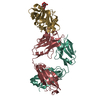
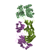
| Title | Crystal structure the Fab fragment of Efalizumab in complex with LFA-1 I domain, Form I | ||||||
 Components Components |
| ||||||
 Keywords Keywords | IMMUNE SYSTEM/CELL ADHESION / Efalizumab / Fab / antibody / LFA-1 / CD11a / I domain / Alternative splicing / Calcium / Glycoprotein / Integrin / Magnesium / Membrane / Polymorphism / Receptor / Transmembrane / IMMUNE SYSTEM-CELL ADHESION COMPLEX | ||||||
| Function / homology |  Function and homology information Function and homology informationmemory T cell extravasation / integrin alphaL-beta2 complex / ICAM-3 receptor activity / T cell activation via T cell receptor contact with antigen bound to MHC molecule on antigen presenting cell / RUNX3 Regulates Immune Response and Cell Migration / heterophilic cell-cell adhesion / integrin complex / leukocyte cell-cell adhesion / receptor clustering / Integrin cell surface interactions ...memory T cell extravasation / integrin alphaL-beta2 complex / ICAM-3 receptor activity / T cell activation via T cell receptor contact with antigen bound to MHC molecule on antigen presenting cell / RUNX3 Regulates Immune Response and Cell Migration / heterophilic cell-cell adhesion / integrin complex / leukocyte cell-cell adhesion / receptor clustering / Integrin cell surface interactions / specific granule membrane / phagocytosis / cell adhesion molecule binding / cell-matrix adhesion / Cell surface interactions at the vascular wall / integrin-mediated signaling pathway / cell-cell adhesion / Immunoregulatory interactions between a Lymphoid and a non-Lymphoid cell / signaling receptor activity / cell adhesion / inflammatory response / Neutrophil degranulation / protein-containing complex binding / cell surface / signal transduction / extracellular exosome / membrane / metal ion binding / plasma membrane Similarity search - Function | ||||||
| Biological species |  Homo sapiens (human) Homo sapiens (human) | ||||||
| Method |  X-RAY DIFFRACTION / SYNCHROTRON / MOLECULAR REPLACEMENT / Resolution: 2.8 Å X-RAY DIFFRACTION / SYNCHROTRON / MOLECULAR REPLACEMENT / Resolution: 2.8 Å | ||||||
 Authors Authors | Li, S. / Ding, J. | ||||||
 Citation Citation | Journal: Proc.Natl.Acad.Sci.USA / Year: 2009 Title: Efalizumab binding to the LFA-1 alphaL I domain blocks ICAM-1 binding via steric hindrance. Authors: Li, S. / Wang, H. / Peng, B. / Zhang, M. / Zhang, D. / Hou, S. / Guo, Y. / Ding, J. | ||||||
| History |
|
- Structure visualization
| Structure viewer | Molecule: MolmilJmol/JSmol |
|---|
- Downloads & links
-Download
| PDBx/mmCIF format | 3eoa.cif.gz | 440.8 KB | Display | PDBx/mmCIF format |
|---|---|---|---|---|
| PDB format | pdb3eoa.ent.gz | 360.1 KB | Display | PDB format |
| PDBx/mmJSON format | 3eoa.json.gz | Tree view | PDBx/mmJSON format | |
| Others |  Other downloads Other downloads |
-Validation report
| Arichive directory | https://data.pdbj.org/pub/pdb/validation_reports/eo/3eoaftp://data.pdbj.org/pub/pdb/validation_reports/eo/3eoa | HTTPS FTP |
|---|
-Related structure data
| Related structure data |  3eo9C  3eobC  1a0qS  1zopS C: citing same article ( S: Starting model for refinement |
|---|---|
| Similar structure data |


-Links
 PDBj
PDBj


- Assembly
| Deposited unit | 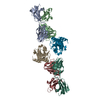
| ||||||||
|---|---|---|---|---|---|---|---|---|---|
| 1 | 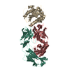
| ||||||||
| 2 | 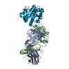
| ||||||||
| Unit cell |
|
-Components
| #1: Antibody | Mass: 23436.068 Da / Num. of mol.: 2 Source method: isolated from a genetically manipulated source Source: (gene. exp.) Homo sapiens (human) / Gene: IGG1#2: Antibody | Mass: 23751.613 Da / Num. of mol.: 2 Source method: isolated from a genetically manipulated source Source: (gene. exp.) Homo sapiens (human) / Gene: IGG1#3: Protein | Mass: 20678.686 Da / Num. of mol.: 2 / Fragment: I domain Source method: isolated from a genetically manipulated source Source: (gene. exp.) Homo sapiens (human) / Gene: ITGAL, CD11A / Plasmid: pET32a / Production host:  #4: Water | ChemComp-HOH / |  Mass: 18.015 Da / Num. of mol.: 356 / Source method: isolated from a natural source / Formula: H2O Mass: 18.015 Da / Num. of mol.: 356 / Source method: isolated from a natural source / Formula: H2OHas protein modification | Y | |
|---|
-Experimental details
-Experiment
| Experiment | Method: X-RAY DIFFRACTION / Number of used crystals: 1 |
|---|
- Sample preparation
| Crystal | Density Matthews: 2.75 Å3/Da / Density % sol: 55.21 % |
|---|---|
| Crystal grow | Temperature: 277 K / Method: vapor diffusion, hanging drop / pH: 5.3 Details: 0.1M sodium citric, 0.2M sodium potassium tartrate, 1.6M ammonium sulfate, pH 5.3, VAPOR DIFFUSION, HANGING DROP, temperature 277K |
-Data collection
| Diffraction | Mean temperature: 100 K |
|---|---|
| Diffraction source | Source: SYNCHROTRON / Site: Photon Factory  / Beamline: AR-NW12A / Wavelength: 1 Å / Beamline: AR-NW12A / Wavelength: 1 Å |
| Detector | Type: ADSC QUANTUM 210 / Detector: CCD / Date: May 24, 2008 / Details: mirrors |
| Radiation | Monochromator: Si 111 CHANNEL / Protocol: SINGLE WAVELENGTH / Monochromatic (M) / Laue (L): M / Scattering type: x-ray |
| Radiation wavelength | Wavelength: 1 Å / Relative weight: 1 |
| Reflection | Resolution: 2.8→50 Å / Num. all: 37885 / Num. obs: 37165 / % possible obs: 98.1 % / Observed criterion σ(F): 1 / Observed criterion σ(I): 1 / Redundancy: 12.5 % / Rmerge(I) obs: 0.118 / Rsym value: 0.118 / Net I/σ(I): 21.1 |
| Reflection shell | Resolution: 2.8→2.9 Å / Redundancy: 4.8 % / Rmerge(I) obs: 0.343 / Mean I/σ(I) obs: 2.7 / Num. unique all: 3139 / Rsym value: 0.343 / % possible all: 84.7 |
- Processing
| Software |
| |||||||||||||||||||||||||||||||||||||||||||||||||||||||||||||||||||||||||||||||||||||||||||||||||||||||||
|---|---|---|---|---|---|---|---|---|---|---|---|---|---|---|---|---|---|---|---|---|---|---|---|---|---|---|---|---|---|---|---|---|---|---|---|---|---|---|---|---|---|---|---|---|---|---|---|---|---|---|---|---|---|---|---|---|---|---|---|---|---|---|---|---|---|---|---|---|---|---|---|---|---|---|---|---|---|---|---|---|---|---|---|---|---|---|---|---|---|---|---|---|---|---|---|---|---|---|---|---|---|---|---|---|---|---|
| Refinement | Method to determine structure: MOLECULAR REPLACEMENT Starting model: 1A0Q, 1ZOP Resolution: 2.8→50 Å / Cor.coef. Fo:Fc: 0.905 / Cor.coef. Fo:Fc free: 0.873 / Cross valid method: THROUGHOUT / σ(F): 1 / σ(I): 1 / ESU R Free: 0.399 / Stereochemistry target values: MAXIMUM LIKELIHOOD
| |||||||||||||||||||||||||||||||||||||||||||||||||||||||||||||||||||||||||||||||||||||||||||||||||||||||||
| Solvent computation | Ion probe radii: 0.8 Å / Shrinkage radii: 0.8 Å / VDW probe radii: 1.2 Å / Solvent model: MASK | |||||||||||||||||||||||||||||||||||||||||||||||||||||||||||||||||||||||||||||||||||||||||||||||||||||||||
| Displacement parameters | Biso mean: 31.434 Å2
| |||||||||||||||||||||||||||||||||||||||||||||||||||||||||||||||||||||||||||||||||||||||||||||||||||||||||
| Refinement step | Cycle: LAST / Resolution: 2.8→50 Å
| |||||||||||||||||||||||||||||||||||||||||||||||||||||||||||||||||||||||||||||||||||||||||||||||||||||||||
| Refine LS restraints |
| |||||||||||||||||||||||||||||||||||||||||||||||||||||||||||||||||||||||||||||||||||||||||||||||||||||||||
| LS refinement shell | Resolution: 2.8→2.873 Å / Total num. of bins used: 20
|
