 Movie
Movie Controller

[English] 日本語
 Yorodumi
- PDB-2y6t: Molecular Recognition of Chymotrypsin by the Serine Protease Inhi... -
+ Open data

- Basic information
| Entry | Database: PDB / ID: 2y6t | ||||||
|---|---|---|---|---|---|---|---|
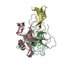
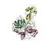
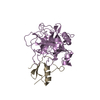
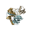
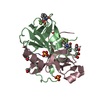
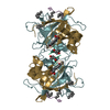
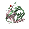
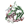
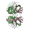
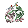
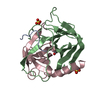
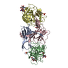
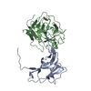
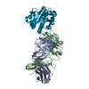
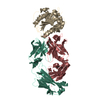
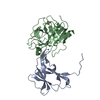
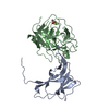
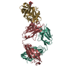
| Title | Molecular Recognition of Chymotrypsin by the Serine Protease Inhibitor Ecotin from Yersinia pestis | ||||||
 Components Components |
| ||||||
 Keywords Keywords | HYDROLASE/INHIBITOR / HYDROLASE-INHIBITOR COMPLEX | ||||||
| Function / homology |  Function and homology information Function and homology informationchymotrypsin / serpin family protein binding / serine protease inhibitor complex / digestion / serine-type endopeptidase inhibitor activity / periplasmic space / serine-type endopeptidase activity / proteolysis / extracellular region Similarity search - Function | ||||||
| Biological species |  YERSINIA PSEUDOTUBERCULOSIS (bacteria) YERSINIA PSEUDOTUBERCULOSIS (bacteria) | ||||||
| Method |  X-RAY DIFFRACTION / SYNCHROTRON / MOLECULAR REPLACEMENT / Resolution: 2.74 Å X-RAY DIFFRACTION / SYNCHROTRON / MOLECULAR REPLACEMENT / Resolution: 2.74 Å | ||||||
 Authors Authors | Clark, E.A. / Walker, N. / Ford, D.C. / Cooper, I.A. / Oyston, P.C.F. / Acharya, K.R. | ||||||
 Citation Citation | Journal: J.Biol.Chem. / Year: 2011 Title: Molecular Recognition of Chymotrypsin by the Serine Protease Inhibitor Ecotin from Yersinia Pestis. Authors: Clark, E.A. / Walker, N. / Ford, D.C. / Cooper, I.A. / Oyston, P.C. / Acharya, K.R. | ||||||
| History |
| ||||||
| Remark 700 | SHEET DETERMINATION METHOD: DSSP THE SHEETS PRESENTED AS "AG" IN EACH CHAIN ON SHEET RECORDS BELOW ... SHEET DETERMINATION METHOD: DSSP THE SHEETS PRESENTED AS "AG" IN EACH CHAIN ON SHEET RECORDS BELOW IS ACTUALLY AN 6-STRANDED BARREL THIS IS REPRESENTED BY A 7-STRANDED SHEET IN WHICH THE FIRST AND LAST STRANDS ARE IDENTICAL. THE SHEETS PRESENTED AS "BB" IN EACH CHAIN ON SHEET RECORDS BELOW IS ACTUALLY AN 6-STRANDED BARREL THIS IS REPRESENTED BY A 7-STRANDED SHEET IN WHICH THE FIRST AND LAST STRANDS ARE IDENTICAL. THE SHEETS PRESENTED AS "CB" IN EACH CHAIN ON SHEET RECORDS BELOW IS ACTUALLY AN 6-STRANDED BARREL THIS IS REPRESENTED BY A 7-STRANDED SHEET IN WHICH THE FIRST AND LAST STRANDS ARE IDENTICAL. THE SHEETS PRESENTED AS "DF" IN EACH CHAIN ON SHEET RECORDS BELOW IS ACTUALLY AN 6-STRANDED BARREL THIS IS REPRESENTED BY A 7-STRANDED SHEET IN WHICH THE FIRST AND LAST STRANDS ARE IDENTICAL. |
- Structure visualization
| Structure viewer | Molecule: MolmilJmol/JSmol |
|---|
- Downloads & links
-Download
| PDBx/mmCIF format | 2y6t.cif.gz | 286.3 KB | Display | PDBx/mmCIF format |
|---|---|---|---|---|
| PDB format | pdb2y6t.ent.gz | 233.1 KB | Display | PDB format |
| PDBx/mmJSON format | 2y6t.json.gz | Tree view | PDBx/mmJSON format | |
| Others |  Other downloads Other downloads |
-Validation report
| Arichive directory | https://data.pdbj.org/pub/pdb/validation_reports/y6/2y6tftp://data.pdbj.org/pub/pdb/validation_reports/y6/2y6t | HTTPS FTP |
|---|
-Related structure data


-Links
 PDBj
PDBj

- Assembly
| Deposited unit | 
| ||||||||
|---|---|---|---|---|---|---|---|---|---|
| 1 | 
| ||||||||
| 2 | 
| ||||||||
| Unit cell |
|
-Components
| #1: Protein | Mass: 25686.037 Da / Num. of mol.: 4 / Source method: isolated from a natural source Details: ACTIVATED BOVINE CHYMOTRYPSIN WAS PURCHASED FROM SIGMA-ALDRICH Source: (natural) #2: Protein | Mass: 16747.180 Da / Num. of mol.: 4 Source method: isolated from a genetically manipulated source Source: (gene. exp.) YERSINIA PSEUDOTUBERCULOSIS (bacteria) / Strain: YPIII / Plasmid: PCRT7/NT-TOPO / Production host: #3: Chemical | ChemComp-SO4 /   Mass: 96.063 Da / Num. of mol.: 4 / Source method: obtained synthetically / Formula: SO4 Mass: 96.063 Da / Num. of mol.: 4 / Source method: obtained synthetically / Formula: SO4#4: Water | ChemComp-HOH / |  Mass: 18.015 Da / Num. of mol.: 33 / Source method: isolated from a natural source / Formula: H2O Mass: 18.015 Da / Num. of mol.: 33 / Source method: isolated from a natural source / Formula: H2OHas protein modification | Y | |
|---|
-Experimental details
-Experiment
| Experiment | Method: X-RAY DIFFRACTION / Number of used crystals: 1 |
|---|
- Sample preparation
| Crystal | Density Matthews: 2.17 Å3/Da / Density % sol: 43.5 % / Description: NONE |
|---|---|
| Crystal grow | pH: 5.5 Details: RESERVOIR SOLUTION - 100 MM BIS TRIS PH 5.5, 200 MM AMMONIUM SULPHATE, 17.5% PEG3350. PROTEIN SOLUTION - 3 MG/ML ECOTIN, 6 MG/ML CHYMOTRYPSIN IN 5MM TRIS PH 7.5. DROP - 1:1 VOLUME RATIO RESERVOIR:PROTEIN. |
-Data collection
| Diffraction | Mean temperature: 100 K |
|---|---|
| Diffraction source | Source: SYNCHROTRON / Site: Diamond  / Beamline: I03 / Wavelength: 0.978 / Beamline: I03 / Wavelength: 0.978 |
| Detector | Type: ADSC CCD / Detector: CCD / Date: Jan 20, 2010 / Details: MIRRORS |
| Radiation | Monochromator: SI (III) / Protocol: SINGLE WAVELENGTH / Monochromatic (M) / Laue (L): M / Scattering type: x-ray |
| Radiation wavelength | Wavelength: 0.978 Å / Relative weight: 1 |
| Reflection | Resolution: 2.74→169.48 Å / Num. obs: 42247 / % possible obs: 97.5 % / Observed criterion σ(I): 0 / Redundancy: 3.2 % / Biso Wilson estimate: 54.6 Å2 / Rmerge(I) obs: 0.08 / Net I/σ(I): 13.27 |
| Reflection shell | Resolution: 2.74→2.84 Å / Redundancy: 3.2 % / Rmerge(I) obs: 0.21 / Mean I/σ(I) obs: 5.51 / % possible all: 96.9 |
- Processing
| Software |
| ||||||||||||||||||||||||||||||||||||||||||||||||||||||||||||||||||||||||||||||||||||||||||||||||||||||||||||||||||||||||||||||||||||||||||||||||||||||||||||||||||||||||||||||||||||||
|---|---|---|---|---|---|---|---|---|---|---|---|---|---|---|---|---|---|---|---|---|---|---|---|---|---|---|---|---|---|---|---|---|---|---|---|---|---|---|---|---|---|---|---|---|---|---|---|---|---|---|---|---|---|---|---|---|---|---|---|---|---|---|---|---|---|---|---|---|---|---|---|---|---|---|---|---|---|---|---|---|---|---|---|---|---|---|---|---|---|---|---|---|---|---|---|---|---|---|---|---|---|---|---|---|---|---|---|---|---|---|---|---|---|---|---|---|---|---|---|---|---|---|---|---|---|---|---|---|---|---|---|---|---|---|---|---|---|---|---|---|---|---|---|---|---|---|---|---|---|---|---|---|---|---|---|---|---|---|---|---|---|---|---|---|---|---|---|---|---|---|---|---|---|---|---|---|---|---|---|---|---|---|---|
| Refinement | Method to determine structure: MOLECULAR REPLACEMENT Starting model: PDB ENTRIES 4CHA AND 1ECZ Resolution: 2.74→169.48 Å / Cor.coef. Fo:Fc: 0.888 / Cor.coef. Fo:Fc free: 0.824 / SU B: 18.243 / SU ML: 0.367 / Cross valid method: THROUGHOUT / ESU R Free: 0.48 / Stereochemistry target values: MAXIMUM LIKELIHOOD / Details: HYDROGENS HAVE BEEN ADDED IN THE RIDING POSITIONS.
| ||||||||||||||||||||||||||||||||||||||||||||||||||||||||||||||||||||||||||||||||||||||||||||||||||||||||||||||||||||||||||||||||||||||||||||||||||||||||||||||||||||||||||||||||||||||
| Solvent computation | Ion probe radii: 0.8 Å / Shrinkage radii: 0.8 Å / VDW probe radii: 1.4 Å / Solvent model: MASK | ||||||||||||||||||||||||||||||||||||||||||||||||||||||||||||||||||||||||||||||||||||||||||||||||||||||||||||||||||||||||||||||||||||||||||||||||||||||||||||||||||||||||||||||||||||||
| Displacement parameters | Biso mean: 35.89 Å2
| ||||||||||||||||||||||||||||||||||||||||||||||||||||||||||||||||||||||||||||||||||||||||||||||||||||||||||||||||||||||||||||||||||||||||||||||||||||||||||||||||||||||||||||||||||||||
| Refinement step | Cycle: LAST / Resolution: 2.74→169.48 Å
| ||||||||||||||||||||||||||||||||||||||||||||||||||||||||||||||||||||||||||||||||||||||||||||||||||||||||||||||||||||||||||||||||||||||||||||||||||||||||||||||||||||||||||||||||||||||
| Refine LS restraints |
|