 Movie
Movie Controller

[English] 日本語
 Yorodumi
- PDB-1afq: CRYSTAL STRUCTURE OF BOVINE GAMMA-CHYMOTRYPSIN COMPLEXED WITH A S... -
+ Open data

- Basic information
| Entry | Database: PDB / ID: 1afq | ||||||
|---|---|---|---|---|---|---|---|
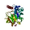
| Title | CRYSTAL STRUCTURE OF BOVINE GAMMA-CHYMOTRYPSIN COMPLEXED WITH A SYNTHETIC INHIBITOR | ||||||
 Components Components | (BOVINE GAMMA- ...) x 3 | ||||||
 Keywords Keywords | HYDROLASE/HYDROLASE INHIBITOR / HYDROLASE-HYDROLASE INHIBITOR COMPLEX | ||||||
| Function / homology |  Function and homology information Function and homology informationchymotrypsin / serpin family protein binding / serine protease inhibitor complex / digestion / serine-type endopeptidase activity / proteolysis / extracellular region Similarity search - Function | ||||||
| Biological species |  | ||||||
| Method |  X-RAY DIFFRACTION / MOLECULAR REPLACEMENT / Resolution: 1.8 Å X-RAY DIFFRACTION / MOLECULAR REPLACEMENT / Resolution: 1.8 Å | ||||||
 Authors Authors | Sugio, S. / Kashima, A. / Inoue, Y. / Maeda, I. / Nose, T. / Shimohigashi, Y. | ||||||
 Citation Citation | Journal: Eur.J.Biochem. / Year: 1998 Title: X-ray crystal structure of a dipeptide-chymotrypsin complex in an inhibitory interaction. Authors: Kashima, A. / Inoue, Y. / Sugio, S. / Maeda, I. / Nose, T. / Shimohigashi, Y. #1: Journal: Biochemistry / Year: 1991Title: Gamma-Chymotrypsin is a Complex of Alpha-Chymotrypsin with its Own Autolysis Products Authors: Harel, M. / Su, C.-T. / Frolow, F. / Silman, I. / Sussman, J.L. #2: Journal: Int.J.Biol.Macromol. / Year: 1991Title: Structure of Gamma-Chymotrypsin in the Range Ph 2.0 To Ph 10.5 Suggests that Gamma-Chymotrypsin is a Covalent Acyl-Enzyme Adduct at Low Ph Authors: Dixon, M.M. / Brennan, R.G. / Matthews, B.W. #3: Journal: Biochemistry / Year: 1989Title: Is Gamma-Chymotrypsin a Tetrapeptide Acyl-Enzyme Adduct of Alpha-Chymotrypsin? Authors: Dixon, M.M. / Matthews, B.W. | ||||||
| History |
|
- Structure visualization
| Structure viewer | Molecule: MolmilJmol/JSmol |
|---|
- Downloads & links
-Download
| PDBx/mmCIF format | 1afq.cif.gz | 61.7 KB | Display | PDBx/mmCIF format |
|---|---|---|---|---|
| PDB format | pdb1afq.ent.gz | 44.1 KB | Display | PDB format |
| PDBx/mmJSON format | 1afq.json.gz | Tree view | PDBx/mmJSON format | |
| Others |  Other downloads Other downloads |
-Validation report
| Arichive directory | https://data.pdbj.org/pub/pdb/validation_reports/af/1afqftp://data.pdbj.org/pub/pdb/validation_reports/af/1afq | HTTPS FTP |
|---|
-Related structure data
| Related structure data |  1ab9SC S: Starting model for refinement C: citing same article ( |
|---|---|
| Similar structure data |


-Links
 PDBj
PDBj

- Assembly
| Deposited unit | 
| |||||||||
|---|---|---|---|---|---|---|---|---|---|---|
| 1 |
| |||||||||
| Unit cell |
| |||||||||
| Components on special symmetry positions |
|
-Components
-BOVINE GAMMA- ... , 3 types, 3 molecules ABC
| #1: Protein/peptide | Mass: 1253.511 Da / Num. of mol.: 1 / Source method: isolated from a natural source / Source: (natural) |
|---|---|
| #2: Protein | Mass: 13934.556 Da / Num. of mol.: 1 / Source method: isolated from a natural source / Source: (natural) |
| #3: Protein | Mass: 10003.417 Da / Num. of mol.: 1 / Source method: isolated from a natural source / Source: (natural) |
-Non-polymers , 3 types, 130 molecules 


| #4: Chemical |  Type: peptide-like, Peptide-like / Class: Inhibitor / Mass: 385.475 Da / Num. of mol.: 2 / Source method: obtained synthetically / Formula: C22H28FN3O2 / Details: AN INHIBITOR FOR CHYMOTRYPSIN / References: D-LEUCYL-L-PHENYLALANYL-P-FLUOROBENZYLAMIDE Type: peptide-like, Peptide-like / Class: Inhibitor / Mass: 385.475 Da / Num. of mol.: 2 / Source method: obtained synthetically / Formula: C22H28FN3O2 / Details: AN INHIBITOR FOR CHYMOTRYPSIN / References: D-LEUCYL-L-PHENYLALANYL-P-FLUOROBENZYLAMIDE#5: Chemical | ChemComp-SO4 / |  Mass: 96.063 Da / Num. of mol.: 1 / Source method: obtained synthetically / Formula: SO4 Mass: 96.063 Da / Num. of mol.: 1 / Source method: obtained synthetically / Formula: SO4#6: Water | ChemComp-HOH / | Mass: 18.015 Da / Num. of mol.: 127 / Source method: isolated from a natural source / Formula: H2O |
|---|
-Details
| Has protein modification | Y |
|---|---|
| Nonpolymer details | THERE ARE TWO INHIBITOR MOLECULES IN AN ASYMMETRIC UNIT. ONE (CHAIN C) IS BOUND TO SUBSTRATE ...THERE ARE TWO INHIBITOR MOLECULES IN AN ASYMMETRIC |
-Experimental details
-Experiment
| Experiment | Method: X-RAY DIFFRACTION / Number of used crystals: 1 |
|---|
- Sample preparation
| Crystal | Density Matthews: 2.29 Å3/Da / Density % sol: 46.18 % | ||||||||||||||||||||||||||||||
|---|---|---|---|---|---|---|---|---|---|---|---|---|---|---|---|---|---|---|---|---|---|---|---|---|---|---|---|---|---|---|---|
| Crystal grow | Temperature: 293 K / pH: 7.5 Details: GAMMA-CHYMOTRYPSIN CRYSTALS WERE SOAKED INTO A SOLUTION CONTAINING SATURATED INHIBITOR, 65% SATURATED AMMONIUM SULFATE AND 100 MM HEPES BUFFER (PH7.5) AT 293K FOR A MONTH. | ||||||||||||||||||||||||||||||
| Crystal grow | *PLUS pH: 5.6 / Method: vapor diffusion | ||||||||||||||||||||||||||||||
| Components of the solutions | *PLUS
|
-Data collection
| Diffraction | Mean temperature: 292 K |
|---|---|
| Diffraction source | Source: ROTATING ANODE / Type: RIGAKU RUH2R / Wavelength: 1.5418 |
| Detector | Type: RIGAKU / Detector: IMAGE PLATE / Date: Nov 10, 1995 / Details: YALE MIRRORS |
| Radiation | Monochromator: NI FILTER / Monochromatic (M) / Laue (L): M / Scattering type: x-ray |
| Radiation wavelength | Wavelength: 1.5418 Å / Relative weight: 1 |
| Reflection | Resolution: 1.8→97.4 Å / Num. obs: 22647 / % possible obs: 98.1 % / Observed criterion σ(I): 1 / Redundancy: 9.5 % / Biso Wilson estimate: 19 Å2 / Rmerge(I) obs: 0.059 / Net I/σ(I): 13.5 |
| Reflection shell | Resolution: 1.8→1.9 Å / Rmerge(I) obs: 0.128 / Mean I/σ(I) obs: 4.09 / % possible all: 95.6 |
| Reflection | *PLUS Num. measured all: 215467 |
| Reflection shell | *PLUS % possible obs: 94.6 % / Rmerge(I) obs: 0.143 / Mean I/σ(I) obs: 4.1 |
- Processing
| Software |
| ||||||||||||||||||||||||||||||||||||||||||||||||||||||||||||||||||||||||||||||||
|---|---|---|---|---|---|---|---|---|---|---|---|---|---|---|---|---|---|---|---|---|---|---|---|---|---|---|---|---|---|---|---|---|---|---|---|---|---|---|---|---|---|---|---|---|---|---|---|---|---|---|---|---|---|---|---|---|---|---|---|---|---|---|---|---|---|---|---|---|---|---|---|---|---|---|---|---|---|---|---|---|---|
| Refinement | Method to determine structure: MOLECULAR REPLACEMENT Starting model: PDB ENTRY 1AB9 Resolution: 1.8→5 Å / Rfactor Rfree error: 0.004 / Data cutoff high absF: 100000 / Data cutoff low absF: 0.1 / Isotropic thermal model: RESTRAINED / Cross valid method: POSTERIORI / σ(F): 2
| ||||||||||||||||||||||||||||||||||||||||||||||||||||||||||||||||||||||||||||||||
| Displacement parameters | Biso mean: 22.4 Å2 | ||||||||||||||||||||||||||||||||||||||||||||||||||||||||||||||||||||||||||||||||
| Refine analyze | Luzzati coordinate error obs: 0.2 Å / Luzzati d res low obs: 5 Å | ||||||||||||||||||||||||||||||||||||||||||||||||||||||||||||||||||||||||||||||||
| Refinement step | Cycle: LAST / Resolution: 1.8→5 Å
| ||||||||||||||||||||||||||||||||||||||||||||||||||||||||||||||||||||||||||||||||
| Refine LS restraints |
| ||||||||||||||||||||||||||||||||||||||||||||||||||||||||||||||||||||||||||||||||
| LS refinement shell | Resolution: 1.8→1.86 Å / Rfactor Rfree error: 0.016 / Total num. of bins used: 10
| ||||||||||||||||||||||||||||||||||||||||||||||||||||||||||||||||||||||||||||||||
| Xplor file |
| ||||||||||||||||||||||||||||||||||||||||||||||||||||||||||||||||||||||||||||||||
| Software | *PLUS Name: X-PLOR / Version: 3.1 / Classification: refinement | ||||||||||||||||||||||||||||||||||||||||||||||||||||||||||||||||||||||||||||||||
| Refinement | *PLUS | ||||||||||||||||||||||||||||||||||||||||||||||||||||||||||||||||||||||||||||||||
| Solvent computation | *PLUS | ||||||||||||||||||||||||||||||||||||||||||||||||||||||||||||||||||||||||||||||||
| Displacement parameters | *PLUS Biso mean: 17.8 Å2 | ||||||||||||||||||||||||||||||||||||||||||||||||||||||||||||||||||||||||||||||||
| Refine LS restraints | *PLUS
| ||||||||||||||||||||||||||||||||||||||||||||||||||||||||||||||||||||||||||||||||
| LS refinement shell | *PLUS Rfactor obs: 0.245 |
