[English] 日本語
Yorodumi
- PDB-5hd9: Crystal Structure of the N-terminal domain of the DNA packaging A... -

+
Open data


ID or keywords:

Loading...

-
Basic information

Entry
Database: PDB / ID: 5hd9
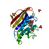
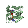
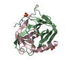
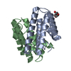
Title
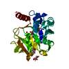
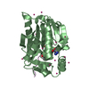
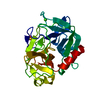
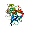
Crystal Structure of the N-terminal domain of the DNA packaging ATPase from bacteriophage phi29
ComponentsEncapsidation protein
KeywordsVIRAL PROTEIN / ASCE fold
Function / homology
Function and homology information


viral DNA genome packaging / Hydrolases; Acting on acid anhydrides; Acting on acid anhydrides to facilitate cellular and subcellular movement / ATP hydrolysis activity / DNA binding / RNA binding / ATP binding
Similarity search - Function
Podovirus DNA packaging protein / Podovirus DNA encapsidation protein (Gp16) / P-loop containing nucleoside triphosphate hydrolase
Similarity search - Domain/homology
DNA packaging protein
Similarity search - Component
Biological speciesBacillus phage phi29 (virus)
MethodX-RAY DIFFRACTION / SYNCHROTRON / SAD / Resolution: 1.941 Å
AuthorsMorais, M.C. / Mao, H. / Reyes-Aldrete, E.
Funding support United States, 2items
OrganizationGrant numberCountry
National Institutes of Health/National Institute of General Medical Sciences (NIH/NIGMS)GM095516 United States
National Institutes of Health/National Institute of General Medical Sciences (NIH/NIGMS)GM059604 United States
CitationJournal: Cell Rep / Year: 2016
Title: Structural and Molecular Basis for Coordination in a Viral DNA Packaging Motor.
Authors: Huzhang Mao / Mitul Saha / Emilio Reyes-Aldrete / Michael B Sherman / Michael Woodson / Rockney Atz / Shelley Grimes / Paul J Jardine / Marc C Morais /
Abstract: Ring NTPases are a class of ubiquitous molecular motors involved in basic biological partitioning processes. dsDNA viruses encode ring ATPases that translocate their genomes to near-crystalline ...Ring NTPases are a class of ubiquitous molecular motors involved in basic biological partitioning processes. dsDNA viruses encode ring ATPases that translocate their genomes to near-crystalline densities within pre-assembled viral capsids. Here, X-ray crystallography, cryoEM, and biochemical analyses of the dsDNA packaging motor in bacteriophage phi29 show how individual subunits are arranged in a pentameric ATPase ring and suggest how their activities are coordinated to translocate dsDNA. The resulting pseudo-atomic structure of the motor and accompanying functional analyses show how ATP is bound in the ATPase active site; identify two DNA contacts, including a potential DNA translocating loop; demonstrate that a trans-acting arginine finger is involved in coordinating hydrolysis around the ring; and suggest a functional coupling between the arginine finger and the DNA translocating loop. The ability to visualize the motor in action illuminates how the different motor components interact with each other and with their DNA substrate.
History
DepositionJan 5, 2016Deposition site: RCSB / Processing site: RCSB
Revision 1.0Mar 9, 2016Provider: repository / Type: Initial release
Revision 1.1Mar 16, 2016Group: Database references
Revision 1.2Sep 13, 2017Group: Author supporting evidence / Derived calculations / Category: pdbx_audit_support / pdbx_struct_oper_list
Item: _pdbx_audit_support.funding_organization / _pdbx_struct_oper_list.symmetry_operation
Revision 1.3Nov 22, 2017Group: Refinement description / Category: software / Item: _software.classification
Revision 1.4Dec 25, 2019Group: Author supporting evidence / Category: pdbx_audit_support / Item: _pdbx_audit_support.funding_organization

-
Structure visualization

Structure viewerMolecule:
MolmilJmol/JSmol

Downloads & links

-
Assembly

Deposited unit
A: Encapsidation protein


Theoretical massNumber of molelcules
Total (without water)22,8551
Polymers22,8551
Non-polymers00
Water3,009167
1


  • Idetical with deposited unit
  • defined by author&software
TypeNameSymmetry operationNumber
identity operation1_555x,y,z1
Buried area0 Å2
ΔGint0 kcal/mol
Surface area10080 Å2
MethodPISA
Unit cell
Length a, b, c (Å)33.103, 36.835, 139.017
Angle α, β, γ (deg.)90.000, 90.000, 90.000
Int Tables number19
Space group name H-MP212121

-
Components

#1: Protein Encapsidation protein / Late protein GP16


Mass: 22854.629 Da / Num. of mol.: 1 / Fragment: N-terminal domain (UNP residues 4-197)
Source method: isolated from a genetically manipulated source
Source: (gene. exp.) Bacillus phage phi29 (virus) / Gene: 16, gp16 / Production host: Escherichia coli (E. coli) / References: UniProt: P11014
#2: Water ChemComp-HOH / water


Mass: 18.015 Da / Num. of mol.: 167 / Source method: isolated from a natural source / Formula: H2O

-
Experimental details

-
Experiment

ExperimentMethod: X-RAY DIFFRACTION / Number of used crystals: 1

-
Sample preparation

CrystalDensity Matthews: 1.85 Å3/Da / Density % sol: 33.66 %
Crystal growTemperature: 290 K / Method: vapor diffusion, sitting drop / pH: 7 / Details: 2.8 M sodium acetate trihydrate

-
Data collection

DiffractionMean temperature: 100 K
Diffraction sourceSource: SYNCHROTRON / Site: CAMD / Beamline: GCPCC / Wavelength: 0.97941 Å
DetectorType: MAR CCD 165 mm / Detector: CCD / Date: Aug 24, 2011
RadiationMonochromator: Si(111) / Protocol: SINGLE WAVELENGTH / Monochromatic (M) / Laue (L): M / Scattering type: x-ray
Radiation wavelengthWavelength: 0.97941 Å / Relative weight: 1
ReflectionResolution: 1.94→35.606 Å / Num. obs: 12985 / % possible obs: 98.19 % / Redundancy: 4.4 % / Net I/σ(I): 31.5

-
Phasing

PhasingMethod: SAD

-
Processing

Software
NameVersionClassification
HKL-2000data collection
SCALEPACKdata scaling
PHENIXrefinement
PDB_EXTRACT3.15data extraction
HKL-2000data reduction
Precognitiondata reduction
RefinementMethod to determine structure: SAD / Resolution: 1.941→35.606 Å / SU ML: 0.17 / Cross valid method: FREE R-VALUE / σ(F): 0 / Phase error: 19.19 / Stereochemistry target values: ML
RfactorNum. reflection% reflection
Rfree0.2092 1294 10 %
Rwork0.1722 11647 -
obs0.1759 12941 97.81 %
Solvent computationShrinkage radii: 0.9 Å / VDW probe radii: 1.11 Å / Solvent model: FLAT BULK SOLVENT MODEL
Displacement parametersBiso max: 80.34 Å2 / Biso mean: 26.1919 Å2 / Biso min: 6.99 Å2
Refinement stepCycle: final / Resolution: 1.941→35.606 Å
ProteinNucleic acidLigandSolventTotal
Num. atoms1606 0 0 167 1773
Biso mean---30.91 -
Num. residues----194
Refine LS restraints
Refine-IDTypeDev idealNumber
X-RAY DIFFRACTIONf_bond_d0.0041648
X-RAY DIFFRACTIONf_angle_d0.9422234
X-RAY DIFFRACTIONf_chiral_restr0.037237
X-RAY DIFFRACTIONf_plane_restr0.004290
X-RAY DIFFRACTIONf_dihedral_angle_d13.844611
LS refinement shell

Refine-ID: X-RAY DIFFRACTION / Total num. of bins used: 9

Resolution (Å)Rfactor RfreeNum. reflection RfreeRfactor RworkNum. reflection RworkNum. reflection all% reflection obs (%)
1.9414-2.01910.251300.19821166129692
2.0191-2.1110.241390.18271260139997
2.111-2.22230.23391400.17111271141198
2.2223-2.36150.21441450.17241303144899
2.3615-2.54380.24691430.191212881431100
2.5438-2.79970.22071450.189513121457100
2.7997-3.20450.21751460.17461313145999
3.2045-4.03650.16841490.15071338148799
4.0365-35.61230.19511570.16711396155397
Refinement TLS params.

Method: refined / Refine-ID: X-RAY DIFFRACTION

IDL112)L122)L132)L222)L232)L332)S11 (Å °)S12 (Å °)S13 (Å °)S21 (Å °)S22 (Å °)S23 (Å °)S31 (Å °)S32 (Å °)S33 (Å °)T112)T122)T132)T222)T232)T332)Origin x (Å)Origin y (Å)Origin z (Å)
17.0820.9582.38382.184-0.04513.248-0.1472-0.01570.06160.06410.0543-0.21610.14460.37920.05940.14160.0067-0.01390.13790.0460.14119.44614.624114.7197
26.3953-2.1399-4.33222.1151.89583.18660.0306-0.0340.1599-0.1708-0.0323-0.0272-0.26480.20630.0360.0965-0.0145-0.01990.1120.00690.18799.951510.255426.2028
31.37730.30270.50213.12570.75753.8849-0.05980.0316-0.2219-0.36060.0682-0.27250.33360.2439-0.01430.12740.00180.0480.09020.02830.15966.0511-2.29921.5591
43.9410.9447-0.30083.8359-0.04763.4922-0.062-0.2497-0.302-0.033-0.0014-0.46830.04690.5826-0.00320.1066-0.01220.0120.20950.03610.194412.14060.781732.0649
53.73040.6060.37082.4272-1.18393.7026-0.03720.1489-0.1154-0.2760.15530.11960.3302-0.221-0.07870.135-0.0257-0.01730.13090.00470.1459-2.39672.674618.5091
67.968-2.79320.94664.9491-1.02846.01880.0860.84380.187-0.604-0.1203-0.12710.4907-0.0492-0.04540.2252-0.02820.00510.21760.03650.10360.58436.64724.3422
78.7090.158-0.56972.88270.14022.171-0.3146-0.04040.0194-0.06780.1373-0.1575-0.22580.37340.17140.1674-0.0443-0.03410.14970.03010.16937.656216.80286.0792
Refinement TLS group
IDRefine-IDRefine TLS-IDSelection detailsAuth asym-IDAuth seq-ID
1X-RAY DIFFRACTION1chain 'A' and (resid 4 through 28 )A0
2X-RAY DIFFRACTION2chain 'A' and (resid 29 through 45 )A0
3X-RAY DIFFRACTION3chain 'A' and (resid 46 through 66 )A0
4X-RAY DIFFRACTION4chain 'A' and (resid 67 through 89 )A0
5X-RAY DIFFRACTION5chain 'A' and (resid 90 through 156 )A0
6X-RAY DIFFRACTION6chain 'A' and (resid 157 through 170 )A0
7X-RAY DIFFRACTION7chain 'A' and (resid 171 through 197 )A0

+
About Yorodumi

-
News

-
Feb 9, 2022. New format data for meta-information of EMDB entries

New format data for meta-information of EMDB entries

  • Version 3 of the EMDB header file is now the official format.
  • The previous official version 1.9 will be removed from the archive.

Related info.:EMDB header

External links:wwPDB to switch to version 3 of the EMDB data model

-
Aug 12, 2020. Covid-19 info

Covid-19 info

URL: https://pdbjlvh1.pdbj.org/emnavi/covid19.php

New page: Covid-19 featured information page in EM Navigator.

Related info.:Covid-19 info / Mar 5, 2020. Novel coronavirus structure data

+
Mar 5, 2020. Novel coronavirus structure data

Novel coronavirus structure data

Related info.:Yorodumi Speices / Aug 12, 2020. Covid-19 info

External links:COVID-19 featured content - PDBj / Molecule of the Month (242):Coronavirus Proteases

+
Jan 31, 2019. EMDB accession codes are about to change! (news from PDBe EMDB page)

EMDB accession codes are about to change! (news from PDBe EMDB page)

  • The allocation of 4 digits for EMDB accession codes will soon come to an end. Whilst these codes will remain in use, new EMDB accession codes will include an additional digit and will expand incrementally as the available range of codes is exhausted. The current 4-digit format prefixed with “EMD-” (i.e. EMD-XXXX) will advance to a 5-digit format (i.e. EMD-XXXXX), and so on. It is currently estimated that the 4-digit codes will be depleted around Spring 2019, at which point the 5-digit format will come into force.
  • The EM Navigator/Yorodumi systems omit the EMD- prefix.

Related info.:Q: What is EMD? / ID/Accession-code notation in Yorodumi/EM Navigator

External links:EMDB Accession Codes are Changing Soon! / Contact to PDBj

+
Jul 12, 2017. Major update of PDB

Major update of PDB

  • wwPDB released updated PDB data conforming to the new PDBx/mmCIF dictionary.
  • This is a major update changing the version number from 4 to 5, and with Remediation, in which all the entries are updated.
  • In this update, many items about electron microscopy experimental information are reorganized (e.g. em_software).
  • Now, EM Navigator and Yorodumi are based on the updated data.

External links:wwPDB Remediation / Enriched Model Files Conforming to OneDep Data Standards Now Available in the PDB FTP Archive

-
Yorodumi

Thousand views of thousand structures

  • Yorodumi is a browser for structure data from EMDB, PDB, SASBDB, etc.
  • This page is also the successor to EM Navigator detail page, and also detail information page/front-end page for Omokage search.
  • The word "yorodu" (or yorozu) is an old Japanese word meaning "ten thousand". "mi" (miru) is to see.

Related info.:EMDB / PDB / SASBDB / Comparison of 3 databanks / Yorodumi Search / Aug 31, 2016. New EM Navigator & Yorodumi / Yorodumi Papers / Jmol/JSmol / Function and homology information / Changes in new EM Navigator and Yorodumi

Read more