 Movie
Movie Controller


+ Open data

- Basic information
| Entry | Database: PDB / ID: 1h0b | ||||||
|---|---|---|---|---|---|---|---|
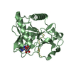
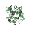
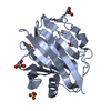
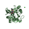
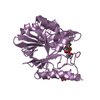
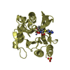
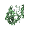
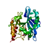
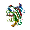
| Title | Endoglucanase cel12A from Rhodothermus marinus | ||||||
 Components Components | CELLULASE | ||||||
 Keywords Keywords | HYDROLASE / CELLULASE / ENDOGLUCANASE | ||||||
| Function / homology |  Function and homology information Function and homology information | ||||||
| Biological species |   RHODOTHERMUS MARINUS (bacteria) RHODOTHERMUS MARINUS (bacteria) | ||||||
| Method |  X-RAY DIFFRACTION / MOLECULAR REPLACEMENT / Resolution: 1.8 Å X-RAY DIFFRACTION / MOLECULAR REPLACEMENT / Resolution: 1.8 Å | ||||||
 Authors Authors | Crennell, S.J. / Hreggvidsson, G.O. / Nordberg-Karlsson, E. | ||||||
 Citation Citation | Journal: J.Mol.Biol. / Year: 2002 Title: The Structure of Rhodothermus Marinus Cel12A, a Highly Thermostable Family 12 Endoglucanase, at 1.8 A Resolution Authors: Crennell, S.J. / Hreggvidsson, G.O. / Nordberg Karlsson, E. | ||||||
| History |
| ||||||
| Remark 700 | SHEET THE SHEET STRUCTURE OF THIS MOLECULE IS BIFURCATED. IN ORDER TO REPRESENT THIS FEATURE IN ... SHEET THE SHEET STRUCTURE OF THIS MOLECULE IS BIFURCATED. IN ORDER TO REPRESENT THIS FEATURE IN THE SHEET RECORDS BELOW, TWO SHEETS ARE DEFINED. |
- Structure visualization
| Structure viewer | Molecule: MolmilJmol/JSmol |
|---|
- Downloads & links
-Download
| PDBx/mmCIF format | 1h0b.cif.gz | 109.5 KB | Display | PDBx/mmCIF format |
|---|---|---|---|---|
| PDB format | pdb1h0b.ent.gz | 85 KB | Display | PDB format |
| PDBx/mmJSON format | 1h0b.json.gz | Tree view | PDBx/mmJSON format | |
| Others |  Other downloads Other downloads |
-Validation report
| Arichive directory | https://data.pdbj.org/pub/pdb/validation_reports/h0/1h0bftp://data.pdbj.org/pub/pdb/validation_reports/h0/1h0b | HTTPS FTP |
|---|
-Related structure data
| Related structure data |  1nlrS S: Starting model for refinement |
|---|---|
| Similar structure data |

-Links
 PDBj
PDBj


- Assembly
| Deposited unit | 
| ||||||||
|---|---|---|---|---|---|---|---|---|---|
| 1 | 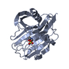
| ||||||||
| 2 | 
| ||||||||
| Unit cell |
| ||||||||
| Noncrystallographic symmetry (NCS) | NCS oper: (Code: given Matrix: (-0.999284, 0.030096, -0.0229), Vector: |
-Components
| #1: Protein | Mass: 28503.469 Da / Num. of mol.: 2 / Fragment: CATALYTIC DOMAIN, RESIDUES 38-260 Source method: isolated from a genetically manipulated source Source: (gene. exp.) RHODOTHERMUS MARINUS (bacteria) / Plasmid: PET25DELTA(SPL) / Production host: #2: Chemical |   Mass: 238.305 Da / Num. of mol.: 2 / Source method: obtained synthetically / Formula: C8H18N2O4S / Comment: pH buffer*YM Mass: 238.305 Da / Num. of mol.: 2 / Source method: obtained synthetically / Formula: C8H18N2O4S / Comment: pH buffer*YM#3: Water | ChemComp-HOH / |  Mass: 18.015 Da / Num. of mol.: 280 / Source method: isolated from a natural source / Formula: H2O Mass: 18.015 Da / Num. of mol.: 280 / Source method: isolated from a natural source / Formula: H2OHas protein modification | Y | Sequence details | CONFLICTS BETWEEN SWISS-PROT ENTRY O33897 AND PDB ENTRY 1H0B ARE AS A RESULT OF RE-SEQUENCING OF ...CONFLICTS BETWEEN SWISS-PROT ENTRY O33897 AND PDB ENTRY 1H0B ARE AS A RESULT OF RE-SEQUENCING | |
|---|
-Experimental details
-Experiment
| Experiment | Method: X-RAY DIFFRACTION / Number of used crystals: 1 |
|---|
- Sample preparation
| Crystal | Density Matthews: 2.49 Å3/Da / Density % sol: 46 % | ||||||||||||||||||||||||
|---|---|---|---|---|---|---|---|---|---|---|---|---|---|---|---|---|---|---|---|---|---|---|---|---|---|
| Crystal grow | pH: 7.5 / Details: 0.1M HEPES, PH7.5, 20% PEG10000, pH 7.50 | ||||||||||||||||||||||||
| Crystal grow | *PLUS Temperature: 291 K / pH: 7.5 / Method: vapor diffusion, hanging drop | ||||||||||||||||||||||||
| Components of the solutions | *PLUS
|
-Data collection
| Diffraction | Mean temperature: 291 K |
|---|---|
| Diffraction source | Source: ROTATING ANODE / Type: RIGAKU RUH3R / Wavelength: 1.5418 |
| Detector | Type: MARRESEARCH / Detector: IMAGE PLATE / Date: Jan 15, 2001 / Details: OSMIC MIRRORS |
| Radiation | Monochromator: NI FILTER / Protocol: SINGLE WAVELENGTH / Monochromatic (M) / Laue (L): M / Scattering type: x-ray |
| Radiation wavelength | Wavelength: 1.5418 Å / Relative weight: 1 |
| Reflection | Resolution: 1.8→30 Å / Num. obs: 44464 / % possible obs: 93.4 % / Redundancy: 6.9 % / Biso Wilson estimate: 17.2 Å2 / Rmerge(I) obs: 0.074 / Net I/σ(I): 27.7 |
| Reflection shell | Resolution: 1.8→1.86 Å / Redundancy: 4.6 % / Rmerge(I) obs: 0.275 / Mean I/σ(I) obs: 5.5 / % possible all: 88.1 |
| Reflection | *PLUS Highest resolution: 1.8 Å / Num. measured all: 308104 / Rmerge(I) obs: 0.074 |
| Reflection shell | *PLUS % possible obs: 88.1 % / Rmerge(I) obs: 0.275 / Mean I/σ(I) obs: 5.3 |
- Processing
| Software |
| ||||||||||||||||||||||||||||||||||||||||||||||||||||||||||||||||||||||||||||||||
|---|---|---|---|---|---|---|---|---|---|---|---|---|---|---|---|---|---|---|---|---|---|---|---|---|---|---|---|---|---|---|---|---|---|---|---|---|---|---|---|---|---|---|---|---|---|---|---|---|---|---|---|---|---|---|---|---|---|---|---|---|---|---|---|---|---|---|---|---|---|---|---|---|---|---|---|---|---|---|---|---|---|
| Refinement | Method to determine structure: MOLECULAR REPLACEMENT Starting model: PDB ENTRY 1NLR Resolution: 1.8→28.49 Å / Rfactor Rfree error: 0.004 / Isotropic thermal model: RESTRAINED / Cross valid method: THROUGHOUT / σ(F): 0
| ||||||||||||||||||||||||||||||||||||||||||||||||||||||||||||||||||||||||||||||||
| Solvent computation | Solvent model: BULK SOLVENT MODEL / Bsol: 38.3243 Å2 / ksol: 0.380923 e/Å3 | ||||||||||||||||||||||||||||||||||||||||||||||||||||||||||||||||||||||||||||||||
| Displacement parameters | Biso mean: 22.38 Å2
| ||||||||||||||||||||||||||||||||||||||||||||||||||||||||||||||||||||||||||||||||
| Refine analyze |
| ||||||||||||||||||||||||||||||||||||||||||||||||||||||||||||||||||||||||||||||||
| Refinement step | Cycle: LAST / Resolution: 1.8→28.49 Å
| ||||||||||||||||||||||||||||||||||||||||||||||||||||||||||||||||||||||||||||||||
| Refine LS restraints |
| ||||||||||||||||||||||||||||||||||||||||||||||||||||||||||||||||||||||||||||||||
| LS refinement shell | Resolution: 1.8→1.88 Å / Rfactor Rfree error: 0.016 / Total num. of bins used: 10
| ||||||||||||||||||||||||||||||||||||||||||||||||||||||||||||||||||||||||||||||||
| Xplor file |
| ||||||||||||||||||||||||||||||||||||||||||||||||||||||||||||||||||||||||||||||||
| Refinement | *PLUS Highest resolution: 1.8 Å / Lowest resolution: 30 Å / Num. reflection obs: 42154 | ||||||||||||||||||||||||||||||||||||||||||||||||||||||||||||||||||||||||||||||||
| Solvent computation | *PLUS | ||||||||||||||||||||||||||||||||||||||||||||||||||||||||||||||||||||||||||||||||
| Displacement parameters | *PLUS | ||||||||||||||||||||||||||||||||||||||||||||||||||||||||||||||||||||||||||||||||
| Refine LS restraints | *PLUS
|
