[English] 日本語
Yorodumi
- PDB-1uu6: X-RAY CRYSTAL STRUCTURE OF THE CATALYTIC DOMAIN OF HUMICOLA GRISE... -

+
Open data


ID or keywords:

Loading...

-
Basic information

Entry
Database: PDB / ID: 1uu6
TitleX-RAY CRYSTAL STRUCTURE OF THE CATALYTIC DOMAIN OF HUMICOLA GRISEA CEL12A IN COMPLEX WITH A SOAKED CELLOPENTAOSE
ComponentsENDO-BETA-1,4-GLUCANASE
KeywordsHYDROLASE / CELLULASE / CELLULOSE DEGRADATION / ENDOGLUCANASE / GLYCOSYL HYDROLASE / GH FAMILY 12 / HUMICOLA GRISEA CEL12A / LIGAND COMPLEX
Function / homology
Function and homology information


cellulase activity / polysaccharide catabolic process
Similarity search - Function
Glycoside hydrolase family 12 / Glycosyl hydrolase family 12 / Glycoside hydrolase family 11/12, catalytic domain / Glycoside hydrolase family 11/12 / Concanavalin A-like lectin/glucanase domain superfamily / Jelly Rolls / Sandwich / Mainly Beta
Similarity search - Domain/homology
beta-cellotetraose / Endoglucanase
Similarity search - Component
Biological speciesHUMICOLA GRISEA (fungus)
MethodX-RAY DIFFRACTION / SYNCHROTRON / OTHER / Resolution: 1.4 Å
AuthorsBerglund, G.I. / Shaw, A. / Stahlberg, J. / Kenne, L. / Driguez, T.H. / Mitchinson, C. / Sandgren, M.
CitationJournal: J.Mol.Biol. / Year: 2004
Title: Crystal Complex Structures Reveal How Substrate is Bound in the -4 to the +2 Binding Sites of Humicola Grisea Cel12A
Authors: Sandgren, M. / Berglund, G.I. / Shaw, A. / Stahlberg, J. / Kenne, L. / Desmet, T. / Mitchinson, C.
History
DepositionDec 15, 2003Deposition site: PDBE / Processing site: PDBE
Revision 1.0Sep 16, 2004Provider: repository / Type: Initial release
Revision 1.1May 8, 2011Group: Version format compliance
Revision 1.2Jul 13, 2011Group: Version format compliance
Revision 2.0Mar 11, 2020Group: Advisory / Data collection ...Advisory / Data collection / Derived calculations / Other / Polymer sequence
Category: chem_comp / entity_poly ...chem_comp / entity_poly / pdbx_database_status / pdbx_unobs_or_zero_occ_atoms / struct_conn
Item: _chem_comp.type / _entity_poly.pdbx_seq_one_letter_code_can ..._chem_comp.type / _entity_poly.pdbx_seq_one_letter_code_can / _pdbx_database_status.status_code_sf / _struct_conn.pdbx_leaving_atom_flag
Revision 3.0Jul 29, 2020Group: Atomic model / Data collection ...Atomic model / Data collection / Derived calculations / Structure summary
Category: atom_site / chem_comp ...atom_site / chem_comp / entity / entity_name_com / pdbx_branch_scheme / pdbx_chem_comp_identifier / pdbx_entity_branch / pdbx_entity_branch_descriptor / pdbx_entity_branch_link / pdbx_entity_branch_list / pdbx_entity_nonpoly / pdbx_molecule_features / pdbx_nonpoly_scheme / pdbx_struct_assembly_gen / struct_asym / struct_conn / struct_site / struct_site_gen
Item: _atom_site.auth_asym_id / _atom_site.auth_seq_id ..._atom_site.auth_asym_id / _atom_site.auth_seq_id / _atom_site.label_asym_id / _chem_comp.name / _entity.formula_weight / _entity.pdbx_description / _entity.pdbx_number_of_molecules / _entity.type / _pdbx_struct_assembly_gen.asym_id_list / _struct_conn.ptnr1_auth_asym_id / _struct_conn.ptnr1_auth_seq_id / _struct_conn.ptnr1_label_asym_id / _struct_conn.ptnr2_auth_asym_id / _struct_conn.ptnr2_auth_seq_id / _struct_conn.ptnr2_label_asym_id
Description: Carbohydrate remediation / Provider: repository / Type: Remediation
Remark 700 
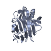
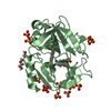
SHEET THE SHEET STRUCTURE OF THIS MOLECULE IS BIFURCATED. IN ORDER TO REPRESENT THIS FEATURE IN ... SHEET THE SHEET STRUCTURE OF THIS MOLECULE IS BIFURCATED. IN ORDER TO REPRESENT THIS FEATURE IN THE SHEET RECORDS BELOW, TWO SHEETS ARE DEFINED.

-
Structure visualization

Structure viewerMolecule:
MolmilJmol/JSmol

Downloads & links

-
Assembly

Deposited unit
A: ENDO-BETA-1,4-GLUCANASE
hetero molecules


Theoretical massNumber of molelcules
Total (without water)26,7493
Polymers25,8891
Non-polymers8612
Water3,747208
1


  • Idetical with deposited unit
  • defined by author&software
TypeNameSymmetry operationNumber
identity operation1_555x,y,z1
MethodPQS
Unit cell
Length a, b, c (Å)49.340, 49.340, 166.061
Angle α, β, γ (deg.)90.00, 90.00, 90.00
Int Tables number96
Space group name H-MP43212

-
Components

#1: Protein ENDO-BETA-1,4-GLUCANASE / ENDOGLUCANASE / CEL12A


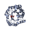
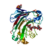
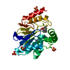
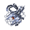
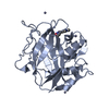
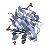
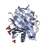
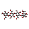
Mass: 25888.586 Da / Num. of mol.: 1 / Fragment: CATALYTIC DOMAIN, RESIDUES 31-254
Source method: isolated from a genetically manipulated source
Details: THE CRYSTAL STRUCTURE IS A COMPLEX WITH A SOAKED CELLOPENTAOSE. THE FIFTH GLUCOSE UNIT IS, HOWEVER, NOT VISIBLE IN THE ELECTRON DENSITY
Source: (gene. exp.) HUMICOLA GRISEA (fungus) / Production host: ASPERGILLUS NIGER (mold) / References: UniProt: Q8NJY3, cellulase
#2: Polysaccharide beta-D-glucopyranose-(1-4)-beta-D-glucopyranose-(1-4)-beta-D-glucopyranose-(1-4)-beta-D-glucopyranose / beta-cellotetraose


Type: oligosaccharide, Oligosaccharide / Class: Metabolism / Mass: 666.578 Da / Num. of mol.: 1
Source method: isolated from a genetically manipulated source
Details: oligosaccharide / References: beta-cellotetraose
DescriptorTypeProgram
DGlcpb1-4DGlcpb1-4DGlcpb1-4DGlcpb1-ROHGlycam Condensed SequenceGMML 1.0
WURCS=2.0/1,4,3/[a2122h-1b_1-5]/1-1-1-1/a4-b1_b4-c1_c4-d1WURCSPDB2Glycan 1.1.0
[][b-D-Glcp]{[(4+1)][b-D-Glcp]{[(4+1)][b-D-Glcp]{[(4+1)][b-D-Glcp]{}}}}LINUCSPDB-CARE
#3: Chemical ChemComp-PG4 / TETRAETHYLENE GLYCOL / Polyethylene glycol


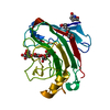
Mass: 194.226 Da / Num. of mol.: 1 / Source method: obtained synthetically / Formula: C8H18O5 / Comment: precipitant*YM
#4: Water ChemComp-HOH / water / Water


Mass: 18.015 Da / Num. of mol.: 208 / Source method: isolated from a natural source / Formula: H2O

-
Experimental details

-
Experiment

ExperimentMethod: X-RAY DIFFRACTION / Number of used crystals: 1

-
Sample preparation

CrystalDensity Matthews: 1.8 Å3/Da / Density % sol: 30.6 %
Description: INITIAL MODEL WAS PRODUCED BY RIGID BODY REFINEMENT USING THE APO PROTEIN STRUCTURE PDB ENTRY 1OLR
Crystal growpH: 3.1
Details: CRYSTALS GREW FROM A PROTEIN STOCK SOLUTION CONTAINING 1MG/ML PROTEIN IN 0.05 M BIS TRIS PROPANE AND 0.05 M AMMONIUM ACETATE, PH 8. CRYSTALS WERE CRYOPROTECTED IN UNBUFFERED 50% MME PEG 2000

-
Data collection

DiffractionMean temperature: 100 K
Diffraction sourceSource: SYNCHROTRON / Site: ESRF / Beamline: ID14-1 / Wavelength: 0.934
DetectorType: ADSC CCD / Detector: CCD / Date: Oct 5, 2001
RadiationProtocol: SINGLE WAVELENGTH / Monochromatic (M) / Laue (L): M / Scattering type: x-ray
Radiation wavelengthWavelength: 0.934 Å / Relative weight: 1
ReflectionResolution: 1.4→42.258 Å / Num. obs: 41450 / % possible obs: 99.7 % / Redundancy: 9.7 % / Rmerge(I) obs: 0.088 / Net I/σ(I): 20.4
Reflection shellResolution: 1.4→1.42 Å / Rmerge(I) obs: 0.316 / Mean I/σ(I) obs: 13.8 / % possible all: 99.2

-
Processing

Software
NameVersionClassification
REFMAC5.1.24refinement
DENZOdata reduction
SCALEPACKdata scaling
RefinementMethod to determine structure: OTHER / Resolution: 1.4→42.26 Å / Cor.coef. Fo:Fc: 0.969 / Cor.coef. Fo:Fc free: 0.964 / Cross valid method: THROUGHOUT / ESU R: 0.054 / ESU R Free: 0.055 / Stereochemistry target values: MAXIMUM LIKELIHOOD
Details: HYDROGENS HAVE BEEN ADDED IN THE RIDING POSITIONS. THE FOLLOWING PROTEIN RESIDUES HAVE BEEN MODELED IN MULTIPLE CONFORMATIONS: A49 A141 A167 A174 A183 A224. THE FOLLOWING WATERS HAVE BEEN ...Details: HYDROGENS HAVE BEEN ADDED IN THE RIDING POSITIONS. THE FOLLOWING PROTEIN RESIDUES HAVE BEEN MODELED IN MULTIPLE CONFORMATIONS: A49 A141 A167 A174 A183 A224. THE FOLLOWING WATERS HAVE BEEN MODELED IN MULTIPLE CONFORMATIONS: W17 W40 W45 W73 W75 W78 W112 W113 W114 W127 W147 W170 W171 W180 W186 ATOMS WITH MISSING ELECTRON DENSITY ARE ASSIGNED ZERO OCCUPANCY. ATOMS ARE ASSIGNED REDUCED OCCUPANCIES WHEN ELECTRON DENSITY IS WEAK OR ATOMS HAVE PARTIAL OCCUPANCY.
RfactorNum. reflection% reflectionSelection details
Rfree0.168 1298 3.1 %RANDOM
Rwork0.147 ---
obs0.148 40151 100 %-
Solvent computationIon probe radii: 0.8 Å / Shrinkage radii: 0.8 Å / VDW probe radii: 1.4 Å / Solvent model: BABINET MODEL WITH MASK
Displacement parametersBiso mean: 8.92 Å2
Baniso -1Baniso -2Baniso -3
1-0 Å20 Å20 Å2
2--0 Å20 Å2
3---0 Å2
Refinement stepCycle: LAST / Resolution: 1.4→42.26 Å
ProteinNucleic acidLigandSolventTotal
Num. atoms1832 0 58 208 2098
Refine LS restraints
Refine-IDTypeDev idealDev ideal targetNumber
X-RAY DIFFRACTIONr_bond_refined_d0.0080.0211984
X-RAY DIFFRACTIONr_bond_other_d00.021642
X-RAY DIFFRACTIONr_angle_refined_deg1.3361.9372717
X-RAY DIFFRACTIONr_angle_other_deg3.79333800
X-RAY DIFFRACTIONr_dihedral_angle_1_deg7.0735236
X-RAY DIFFRACTIONr_dihedral_angle_2_deg
X-RAY DIFFRACTIONr_dihedral_angle_3_deg
X-RAY DIFFRACTIONr_dihedral_angle_4_deg
X-RAY DIFFRACTIONr_chiral_restr0.0860.2278
X-RAY DIFFRACTIONr_gen_planes_refined0.0060.022219
X-RAY DIFFRACTIONr_gen_planes_other0.0080.02457
X-RAY DIFFRACTIONr_nbd_refined0.1790.2299
X-RAY DIFFRACTIONr_nbd_other0.2780.21893
X-RAY DIFFRACTIONr_nbtor_refined
X-RAY DIFFRACTIONr_nbtor_other0.1120.2936
X-RAY DIFFRACTIONr_xyhbond_nbd_refined0.0960.2114
X-RAY DIFFRACTIONr_xyhbond_nbd_other
X-RAY DIFFRACTIONr_metal_ion_refined
X-RAY DIFFRACTIONr_metal_ion_other
X-RAY DIFFRACTIONr_symmetry_vdw_refined0.1430.214
X-RAY DIFFRACTIONr_symmetry_vdw_other0.3040.273
X-RAY DIFFRACTIONr_symmetry_hbond_refined0.1160.211
X-RAY DIFFRACTIONr_symmetry_hbond_other
X-RAY DIFFRACTIONr_symmetry_metal_ion_refined
X-RAY DIFFRACTIONr_symmetry_metal_ion_other
X-RAY DIFFRACTIONr_mcbond_it0.61.51130
X-RAY DIFFRACTIONr_mcbond_other
X-RAY DIFFRACTIONr_mcangle_it1.16121817
X-RAY DIFFRACTIONr_mcangle_other
X-RAY DIFFRACTIONr_scbond_it1.6243854
X-RAY DIFFRACTIONr_scbond_other
X-RAY DIFFRACTIONr_scangle_it2.4624.5894
X-RAY DIFFRACTIONr_scangle_other
X-RAY DIFFRACTIONr_long_range_B_refined
X-RAY DIFFRACTIONr_long_range_B_other
X-RAY DIFFRACTIONr_rigid_bond_restr
X-RAY DIFFRACTIONr_sphericity_free
X-RAY DIFFRACTIONr_sphericity_bonded
LS refinement shellResolution: 1.4→1.44 Å / Total num. of bins used: 20 /
RfactorNum. reflection
Rfree0.192 89
Rwork0.154 2881

+
About Yorodumi

-
News

-
Feb 9, 2022. New format data for meta-information of EMDB entries

New format data for meta-information of EMDB entries

  • Version 3 of the EMDB header file is now the official format.
  • The previous official version 1.9 will be removed from the archive.

Related info.:EMDB header

External links:wwPDB to switch to version 3 of the EMDB data model

-
Aug 12, 2020. Covid-19 info

Covid-19 info

URL: https://pdbj.org/emnavi/covid19.php

New page: Covid-19 featured information page in EM Navigator.

Related info.:Covid-19 info / Mar 5, 2020. Novel coronavirus structure data

+
Mar 5, 2020. Novel coronavirus structure data

Novel coronavirus structure data

Related info.:Yorodumi Speices / Aug 12, 2020. Covid-19 info

External links:COVID-19 featured content - PDBj / Molecule of the Month (242):Coronavirus Proteases

+
Jan 31, 2019. EMDB accession codes are about to change! (news from PDBe EMDB page)

EMDB accession codes are about to change! (news from PDBe EMDB page)

  • The allocation of 4 digits for EMDB accession codes will soon come to an end. Whilst these codes will remain in use, new EMDB accession codes will include an additional digit and will expand incrementally as the available range of codes is exhausted. The current 4-digit format prefixed with “EMD-” (i.e. EMD-XXXX) will advance to a 5-digit format (i.e. EMD-XXXXX), and so on. It is currently estimated that the 4-digit codes will be depleted around Spring 2019, at which point the 5-digit format will come into force.
  • The EM Navigator/Yorodumi systems omit the EMD- prefix.

Related info.:Q: What is EMD? / ID/Accession-code notation in Yorodumi/EM Navigator

External links:EMDB Accession Codes are Changing Soon! / Contact to PDBj

+
Jul 12, 2017. Major update of PDB

Major update of PDB

  • wwPDB released updated PDB data conforming to the new PDBx/mmCIF dictionary.
  • This is a major update changing the version number from 4 to 5, and with Remediation, in which all the entries are updated.
  • In this update, many items about electron microscopy experimental information are reorganized (e.g. em_software).
  • Now, EM Navigator and Yorodumi are based on the updated data.

External links:wwPDB Remediation / Enriched Model Files Conforming to OneDep Data Standards Now Available in the PDB FTP Archive

-
Yorodumi

Thousand views of thousand structures

  • Yorodumi is a browser for structure data from EMDB, PDB, SASBDB, etc.
  • This page is also the successor to EM Navigator detail page, and also detail information page/front-end page for Omokage search.
  • The word "yorodu" (or yorozu) is an old Japanese word meaning "ten thousand". "mi" (miru) is to see.

Related info.:EMDB / PDB / SASBDB / Comparison of 3 databanks / Yorodumi Search / Aug 31, 2016. New EM Navigator & Yorodumi / Yorodumi Papers / Jmol/JSmol / Function and homology information / Changes in new EM Navigator and Yorodumi

Read more