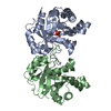
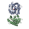
 Movie
Movie Controller


+ Open data

- Basic information
| Entry | Database: PDB / ID: 1nlr | ||||||
|---|---|---|---|---|---|---|---|
| Title | ENDO-1,4-BETA-GLUCANASE CELB2, CELLULASE, NATIVE STRUCTURE | ||||||
 Components Components | ENDO-1,4-BETA-GLUCANASE | ||||||
 Keywords Keywords | ENDOGLUCANASE / GLYCOSYL HYDROLASE / FAMILY 12 / CELB2 | ||||||
| Function / homology |  Function and homology information Function and homology informationcellulase activity / polysaccharide binding / polysaccharide catabolic process Similarity search - Function | ||||||
| Biological species |  Streptomyces lividans (bacteria) Streptomyces lividans (bacteria) | ||||||
| Method |  X-RAY DIFFRACTION / MIR / Resolution: 1.75 Å X-RAY DIFFRACTION / MIR / Resolution: 1.75 Å | ||||||
 Authors Authors | Sulzenbacher, G. / Dupont, C. / Davies, G.J. | ||||||
 Citation Citation | Journal: Biochemistry / Year: 1997 Title: The Streptomyces lividans family 12 endoglucanase: construction of the catalytic cre, expression, and X-ray structure at 1.75 A resolution. Authors: Sulzenbacher, G. / Shareck, F. / Morosoli, R. / Dupont, C. / Davies, G.J. #1: Journal: Nature / Year: 1997Title: Erratum. Structure of the Inhibitory Receptor for Human Natural Killer Cells Resembles Haematopoietic Receptors Authors: Fan, Q.R. / Mosyak, L. / Winter, C.C. / Wagtmann, N. / Long, E.O. / Wiley, D.C. #2: Journal: Appl.Environ.Microbiol. / Year: 1994Title: Purification and Characterization of the Celb Endoglucanase from Streptomyces Lividans 66 and DNA Sequence of the Encoding Gene Authors: Wittmann, S. / Shareck, F. / Kluepfel, D. / Morosoli, R. | ||||||
| History |
|
- Structure visualization
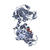
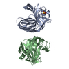
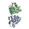
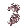
| Structure viewer | Molecule: MolmilJmol/JSmol |
|---|
- Downloads & links
-Download
| PDBx/mmCIF format | 1nlr.cif.gz | 60.7 KB | Display | PDBx/mmCIF format |
|---|---|---|---|---|
| PDB format | pdb1nlr.ent.gz | 43.8 KB | Display | PDB format |
| PDBx/mmJSON format | 1nlr.json.gz | Tree view | PDBx/mmJSON format | |
| Others |  Other downloads Other downloads |
-Validation report
| Arichive directory | https://data.pdbj.org/pub/pdb/validation_reports/nl/1nlrftp://data.pdbj.org/pub/pdb/validation_reports/nl/1nlr | HTTPS FTP |
|---|
-Related structure data
| Similar structure data |
|---|


-Links
 PDBj
PDBj

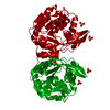
- Assembly
| Deposited unit | 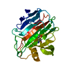
| ||||||||
|---|---|---|---|---|---|---|---|---|---|
| 1 | 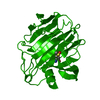
| ||||||||
| Unit cell |
|
-Components
| #1: Protein | Mass: 24594.713 Da / Num. of mol.: 1 / Fragment: CATALYTIC DOMAIN Source method: isolated from a genetically manipulated source Source: (gene. exp.) Streptomyces lividans (bacteria) / Fragment: CATALYTIC DOMAIN / Production host: Streptomyces lividans (bacteria) / Strain (production host): 66 / References: UniProt: Q54331, cellulase |
|---|---|
| #2: Water | ChemComp-HOH /  Mass: 18.015 Da / Num. of mol.: 200 / Source method: isolated from a natural source / Formula: H2O Mass: 18.015 Da / Num. of mol.: 200 / Source method: isolated from a natural source / Formula: H2O |
| Compound details | THE COORDINATES GIVEN DEFINE THE STRUCTURE OF CELB2, THE TRUNCATED, CATALYTICALLY COMPETENT, FORM ...THE COORDINATE |
| Has protein modification | Y |
-Experimental details
-Experiment
| Experiment | Method: X-RAY DIFFRACTION / Number of used crystals: 1 |
|---|
- Sample preparation
| Crystal | Density Matthews: 2.01 Å3/Da / Density % sol: 38.2 % | ||||||||||||||||||||
|---|---|---|---|---|---|---|---|---|---|---|---|---|---|---|---|---|---|---|---|---|---|
| Crystal grow | Method: vapor diffusion / pH: 4.5 Details: 30 % PEG 1500, PH 4.5 FOR ACETATE BUFFER METHOD: HANGING DROP VAPOUR DIFFUSION, vapor diffusion | ||||||||||||||||||||
| Crystal grow | *PLUS Method: vapor diffusion, hanging drop | ||||||||||||||||||||
| Components of the solutions | *PLUS
|
-Data collection
| Diffraction | Mean temperature: 120 K |
|---|---|
| Diffraction source | Source: ROTATING ANODE / Type: RIGAKU FR-C / Wavelength: 1.5418 |
| Detector | Type: RIGAKU RAXIS IIC / Detector: IMAGE PLATE / Date: Feb 17, 1997 / Details: FOCUSING MIRRORS |
| Radiation | Monochromatic (M) / Laue (L): M / Scattering type: x-ray |
| Radiation wavelength | Wavelength: 1.5418 Å / Relative weight: 1 |
| Reflection | Resolution: 1.75→15 Å / Num. obs: 18747 / % possible obs: 95.6 % / Redundancy: 6.98 % / Biso Wilson estimate: 18.51 Å2 / Rmerge(I) obs: 0.047 / Net I/σ(I): 31.4 |
| Reflection shell | Resolution: 1.75→1.78 Å / Redundancy: 3.21 % / Rmerge(I) obs: 0.245 / Mean I/σ(I) obs: 5.96 / % possible all: 68.5 |
| Reflection | *PLUS % possible obs: 93.7 % / Redundancy: 6.77 % |
| Reflection shell | *PLUS Lowest resolution: 1.84 Å / % possible obs: 67.9 % / Redundancy: 2.78 % / Rmerge(I) obs: 0.287 |
- Processing
| Software |
| ||||||||||||||||||||||||||||||||||||||||||||||||||||||||||||||||||||||||||||||||||||
|---|---|---|---|---|---|---|---|---|---|---|---|---|---|---|---|---|---|---|---|---|---|---|---|---|---|---|---|---|---|---|---|---|---|---|---|---|---|---|---|---|---|---|---|---|---|---|---|---|---|---|---|---|---|---|---|---|---|---|---|---|---|---|---|---|---|---|---|---|---|---|---|---|---|---|---|---|---|---|---|---|---|---|---|---|---|
| Refinement | Method to determine structure: MIR / Resolution: 1.75→15 Å / Cross valid method: FREE R
| ||||||||||||||||||||||||||||||||||||||||||||||||||||||||||||||||||||||||||||||||||||
| Displacement parameters | Biso mean: 20.4 Å2 | ||||||||||||||||||||||||||||||||||||||||||||||||||||||||||||||||||||||||||||||||||||
| Refinement step | Cycle: LAST / Resolution: 1.75→15 Å
| ||||||||||||||||||||||||||||||||||||||||||||||||||||||||||||||||||||||||||||||||||||
| Refine LS restraints |
| ||||||||||||||||||||||||||||||||||||||||||||||||||||||||||||||||||||||||||||||||||||
| Software | *PLUS Name: REFMAC / Classification: refinement | ||||||||||||||||||||||||||||||||||||||||||||||||||||||||||||||||||||||||||||||||||||
| Refinement | *PLUS Rfactor obs: 0.187 | ||||||||||||||||||||||||||||||||||||||||||||||||||||||||||||||||||||||||||||||||||||
| Solvent computation | *PLUS | ||||||||||||||||||||||||||||||||||||||||||||||||||||||||||||||||||||||||||||||||||||
| Displacement parameters | *PLUS |
