[English] 日本語
Yorodumi
- PDB-1oxg: Crystal structure of a complex formed between organic solvent tre... -

+
Open data


ID or keywords:

Loading...

-
Basic information

Entry
Database: PDB / ID: 1oxg
Title
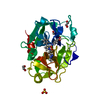
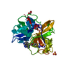
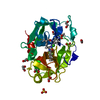
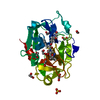
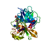
Crystal structure of a complex formed between organic solvent treated bovine alpha-chymotrypsin and its autocatalytically produced highly potent 14-residue peptide at 2.2 resolution
Components(Chymotrypsinogen A) x 2
KeywordsHYDROLASE / Autocatalysis / Organic solvent treatment / Inhibition
Function / homology
Function and homology information


chymotrypsin / serpin family protein binding / serine protease inhibitor complex / digestion / serine-type endopeptidase activity / proteolysis / extracellular region
Similarity search - Function
Serine proteases, trypsin family, histidine active site / Serine proteases, trypsin family, serine active site / Peptidase S1A, chymotrypsin family / Serine proteases, trypsin family, histidine active site. / Serine proteases, trypsin domain profile. / Serine proteases, trypsin family, serine active site. / Trypsin-like serine protease / Serine proteases, trypsin domain / Trypsin / Trypsin-like serine proteases ...Serine proteases, trypsin family, histidine active site / Serine proteases, trypsin family, serine active site / Peptidase S1A, chymotrypsin family / Serine proteases, trypsin family, histidine active site. / Serine proteases, trypsin domain profile. / Serine proteases, trypsin family, serine active site. / Trypsin-like serine protease / Serine proteases, trypsin domain / Trypsin / Trypsin-like serine proteases / Thrombin, subunit H / Peptidase S1, PA clan, chymotrypsin-like fold / Peptidase S1, PA clan / Beta Barrel / Mainly Beta
Similarity search - Domain/homology
Biological speciesBos taurus (cattle)
MethodX-RAY DIFFRACTION / SYNCHROTRON / MOLECULAR REPLACEMENT / Resolution: 2.2 Å
AuthorsSingh, N. / Jabeen, T. / Sharma, S. / Roy, I. / Gupta, M.N. / Bilgrami, S. / Singh, T.P.
CitationJournal: Febs J. / Year: 2005
Title: Detection of native peptides as potent inhibitors of enzymes. Crystal structure of the complex formed between treated bovine alpha-chymotrypsin and an autocatalytically produced fragment, IIe- ...Title: Detection of native peptides as potent inhibitors of enzymes. Crystal structure of the complex formed between treated bovine alpha-chymotrypsin and an autocatalytically produced fragment, IIe-Val-Asn-Gly-Glu-Glu-Ala-Val-Pro-Gly-Ser-Trp-Pro-Trp, at 2.2 angstroms resolution.
Authors: Singh, N. / Jabeen, T. / Sharma, S. / Roy, I. / Gupta, M.N. / Bilgrami, S. / Somvanshi, R.K. / Dey, S. / Perbandt, M. / Betzel, C. / Srinivasan, A. / Singh, T.P.
History
DepositionApr 2, 2003Deposition site: RCSB / Processing site: PDBJ
Revision 1.0May 18, 2004Provider: repository / Type: Initial release
Revision 1.1Apr 29, 2008Group: Version format compliance
Revision 1.2Jul 13, 2011Group: Version format compliance
Revision 2.0Oct 25, 2023Group: Atomic model / Data collection ...Atomic model / Data collection / Database references / Derived calculations / Refinement description
Category: atom_site / chem_comp_atom ...atom_site / chem_comp_atom / chem_comp_bond / database_2 / diffrn_source / pdbx_initial_refinement_model / pdbx_struct_special_symmetry / struct_site
Item: _atom_site.occupancy / _database_2.pdbx_DOI ..._atom_site.occupancy / _database_2.pdbx_DOI / _database_2.pdbx_database_accession / _diffrn_source.pdbx_synchrotron_site / _struct_site.pdbx_auth_asym_id / _struct_site.pdbx_auth_comp_id / _struct_site.pdbx_auth_seq_id

-
Structure visualization

Structure viewerMolecule:
MolmilJmol/JSmol

Downloads & links

-
Assembly

Deposited unit
A: Chymotrypsinogen A
B: Chymotrypsinogen A
hetero molecules


Theoretical massNumber of molelcules
Total (without water)27,8038
Polymers27,2272
Non-polymers5766
Water4,143230
1


  • Idetical with deposited unit
  • defined by author&software
TypeNameSymmetry operationNumber
identity operation1_555x,y,z1
Buried area1510 Å2
ΔGint-53 kcal/mol
Surface area10830 Å2
MethodPISA
Unit cell
Length a, b, c (Å)56.187, 76.382, 105.098
Angle α, β, γ (deg.)90.00, 90.00, 90.00
Int Tables number23
Space group name H-MI222
Components on special symmetry positions
IDModelComponents
11B-1006-

SO4

-
Components

#1: Protein Chymotrypsinogen A / Alpha Chymotrypsin


Mass: 25686.037 Da / Num. of mol.: 1 / Source method: isolated from a natural source / Source: (natural) Bos taurus (cattle) / Secretion: Pancreatic secretion / References: UniProt: P00766, chymotrypsin
#2: Protein/peptide Chymotrypsinogen A / Alpha Chymotrypsin


Mass: 1540.673 Da / Num. of mol.: 1 / Fragment: residues 1-14 / Source method: isolated from a natural source / Source: (natural) Bos taurus (cattle) / Secretion: Pancreatic secretion / References: UniProt: P00766, chymotrypsin
#3: Chemical
ChemComp-SO4 / SULFATE ION


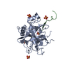
Mass: 96.063 Da / Num. of mol.: 6 / Source method: obtained synthetically / Formula: SO4
#4: Water ChemComp-HOH / water


Mass: 18.015 Da / Num. of mol.: 230 / Source method: isolated from a natural source / Formula: H2O

-
Experimental details

-
Experiment

ExperimentMethod: X-RAY DIFFRACTION / Number of used crystals: 1

-
Sample preparation

CrystalDensity Matthews: 2.07 Å3/Da / Density % sol: 40.6 %
Crystal growTemperature: 298 K / Method: vapor diffusion, sitting drop / pH: 4.8
Details: 0.2M Ammonium sulphate, 20mM Sodium acetate buffer, pH 4.8, VAPOR DIFFUSION, SITTING DROP, temperature 298K

-
Data collection

DiffractionMean temperature: 278 K
Diffraction sourceSource: SYNCHROTRON / Site: EMBL/DESY, HAMBURG / Beamline: X11 / Wavelength: 0.91 Å
DetectorType: MARRESEARCH / Detector: IMAGE PLATE / Date: Apr 14, 2002 / Details: Mirror
RadiationMonochromator: Mirror / Protocol: SINGLE WAVELENGTH / Monochromatic (M) / Laue (L): M / Scattering type: x-ray
Radiation wavelengthWavelength: 0.91 Å / Relative weight: 1
ReflectionResolution: 2.2→20 Å / Num. all: 11726 / Num. obs: 11673 / % possible obs: 98.7 % / Redundancy: 20.3 % / Biso Wilson estimate: 12.3 Å2 / Rsym value: 0.043 / Net I/σ(I): 18.4
Reflection shellResolution: 2.2→2.24 Å / Redundancy: 23.9 % / Mean I/σ(I) obs: 2.9 / Num. unique all: 564 / Rsym value: 0.093 / % possible all: 98.7

-
Processing

Software
NameVersionClassification
CNS0.9refinement
DENZOdata reduction
SCALEPACKdata scaling
AMoREphasing
RefinementMethod to determine structure: MOLECULAR REPLACEMENT
Starting model: 1ACB
Resolution: 2.2→19.69 Å / Rfactor Rfree error: 0.008 / Data cutoff high absF: 1842156.11 / Data cutoff low absF: 0 / Isotropic thermal model: RESTRAINED / Cross valid method: THROUGHOUT / σ(F): 0 / σ(I): 0 / Stereochemistry target values: Engh & Huber
RfactorNum. reflection% reflectionSelection details
Rfree0.207 602 5.2 %RANDOM
Rwork0.192 ---
all0.207 11726 --
obs0.198 11673 98.8 %-
Solvent computationSolvent model: FLAT MODEL / Bsol: 62.6449 Å2 / ksol: 0.342322 e/Å3
Displacement parametersBiso mean: 21.6 Å2
Baniso -1Baniso -2Baniso -3
1--2.91 Å20 Å20 Å2
2--5.23 Å20 Å2
3----2.32 Å2
Refine analyze
FreeObs
Luzzati coordinate error0.24 Å0.21 Å
Luzzati d res low-5 Å
Luzzati sigma a0.16 Å0.07 Å
Refinement stepCycle: LAST / Resolution: 2.2→19.69 Å
ProteinNucleic acidLigandSolventTotal
Num. atoms1855 0 30 230 2115
Refine LS restraints
Refine-IDTypeDev idealDev ideal target
X-RAY DIFFRACTIONc_bond_d0.018
X-RAY DIFFRACTIONc_angle_deg2.3
X-RAY DIFFRACTIONc_dihedral_angle_d27.8
X-RAY DIFFRACTIONc_improper_angle_d1.88
X-RAY DIFFRACTIONc_mcbond_it1.361.5
X-RAY DIFFRACTIONc_mcangle_it2.112
X-RAY DIFFRACTIONc_scbond_it2.192
X-RAY DIFFRACTIONc_scangle_it2.862.5
LS refinement shellResolution: 2.2→2.34 Å / Rfactor Rfree error: 0.026 / Total num. of bins used: 6
RfactorNum. reflection% reflection
Rfree0.239 84 4.4 %
Rwork0.186 1831 -
obs-280936 99.5 %
Xplor file
Refine-IDSerial noParam fileTopol file
X-RAY DIFFRACTION1PROTEIN_REP.PARAMPROTEIN.TOP
X-RAY DIFFRACTION2WATER_REP.PARAMATER.TOP
X-RAY DIFFRACTION3ION.PARAMION.TOP
X-RAY DIFFRACTION4SUL.PARAMSUL.TOP

+
About Yorodumi

-
News

-
Feb 9, 2022. New format data for meta-information of EMDB entries

New format data for meta-information of EMDB entries

  • Version 3 of the EMDB header file is now the official format.
  • The previous official version 1.9 will be removed from the archive.

Related info.:EMDB header

External links:wwPDB to switch to version 3 of the EMDB data model

-
Aug 12, 2020. Covid-19 info

Covid-19 info

URL: https://pdbjlvh1.pdbj.org/emnavi/covid19.php

New page: Covid-19 featured information page in EM Navigator.

Related info.:Covid-19 info / Mar 5, 2020. Novel coronavirus structure data

+
Mar 5, 2020. Novel coronavirus structure data

Novel coronavirus structure data

Related info.:Yorodumi Speices / Aug 12, 2020. Covid-19 info

External links:COVID-19 featured content - PDBj / Molecule of the Month (242):Coronavirus Proteases

+
Jan 31, 2019. EMDB accession codes are about to change! (news from PDBe EMDB page)

EMDB accession codes are about to change! (news from PDBe EMDB page)

  • The allocation of 4 digits for EMDB accession codes will soon come to an end. Whilst these codes will remain in use, new EMDB accession codes will include an additional digit and will expand incrementally as the available range of codes is exhausted. The current 4-digit format prefixed with “EMD-” (i.e. EMD-XXXX) will advance to a 5-digit format (i.e. EMD-XXXXX), and so on. It is currently estimated that the 4-digit codes will be depleted around Spring 2019, at which point the 5-digit format will come into force.
  • The EM Navigator/Yorodumi systems omit the EMD- prefix.

Related info.:Q: What is EMD? / ID/Accession-code notation in Yorodumi/EM Navigator

External links:EMDB Accession Codes are Changing Soon! / Contact to PDBj

+
Jul 12, 2017. Major update of PDB

Major update of PDB

  • wwPDB released updated PDB data conforming to the new PDBx/mmCIF dictionary.
  • This is a major update changing the version number from 4 to 5, and with Remediation, in which all the entries are updated.
  • In this update, many items about electron microscopy experimental information are reorganized (e.g. em_software).
  • Now, EM Navigator and Yorodumi are based on the updated data.

External links:wwPDB Remediation / Enriched Model Files Conforming to OneDep Data Standards Now Available in the PDB FTP Archive

-
Yorodumi

Thousand views of thousand structures

  • Yorodumi is a browser for structure data from EMDB, PDB, SASBDB, etc.
  • This page is also the successor to EM Navigator detail page, and also detail information page/front-end page for Omokage search.
  • The word "yorodu" (or yorozu) is an old Japanese word meaning "ten thousand". "mi" (miru) is to see.

Related info.:EMDB / PDB / SASBDB / Comparison of 3 databanks / Yorodumi Search / Aug 31, 2016. New EM Navigator & Yorodumi / Yorodumi Papers / Jmol/JSmol / Function and homology information / Changes in new EM Navigator and Yorodumi

Read more