 Movie
Movie Controller

[English] 日本語
 Yorodumi
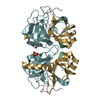
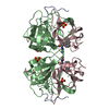
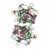
- PDB-2gct: STRUCTURE OF GAMMA-CHYMOTRYPSIN IN THE RANGE PH 2.0 TO PH 10.5 SU... -
+ Open data

- Basic information
| Entry | Database: PDB / ID: 2gct | ||||||
|---|---|---|---|---|---|---|---|
| Title | STRUCTURE OF GAMMA-CHYMOTRYPSIN IN THE RANGE PH 2.0 TO PH 10.5 SUGGESTS THAT GAMMA-CHYMOTRYPSIN IS A COVALENT ACYL-ENZYME ADDUCT AT LOW PH | ||||||
 Components Components |
| ||||||
 Keywords Keywords | HYDROLASE/PEPTIDE / HYDROLASE / SERINE PROTEINASE / HYDROLASE-PEPTIDE COMPLEX | ||||||
| Function / homology |  Function and homology information Function and homology informationchymotrypsin / serpin family protein binding / serine protease inhibitor complex / digestion / serine-type endopeptidase activity / proteolysis / extracellular region Similarity search - Function | ||||||
| Biological species |  | ||||||
| Method |  X-RAY DIFFRACTION / MOLECULAR REPLACEMENT / Resolution: 1.8 Å X-RAY DIFFRACTION / MOLECULAR REPLACEMENT / Resolution: 1.8 Å | ||||||
 Authors Authors | Dixon, M.M. / Matthews, B.W. | ||||||
 Citation Citation | Journal: Int.J.Biol.Macromol. / Year: 1991 Title: Structure of gamma-chymotrypsin in the range pH 2.0 to pH 10.5 suggests that gamma-chymotrypsin is a covalent acyl-enzyme adduct at low pH. Authors: Dixon, M.M. / Brennan, R.G. / Matthews, B.W. #1: Journal: Biochemistry / Year: 1989Title: Is Gamma-Chymotrypsin a Tetrapeptide Acyl-Enzyme Adduct of Gamma-Chymotrypsin? Authors: Dixon, M.M. / Matthews, B.W. | ||||||
| History |
| ||||||
| Remark 700 | SHEET THE SHEET PRESENTED AS *S1* ON SHEET RECORDS BELOW IS ACTUALLY A SIX-STRANDED BETA-BARREL. ...SHEET THE SHEET PRESENTED AS *S1* ON SHEET RECORDS BELOW IS ACTUALLY A SIX-STRANDED BETA-BARREL. THIS IS REPRESENTED BY A SEVEN-STRANDED SHEET IN WHICH THE FIRST AND LAST STRANDS ARE IDENTICAL. SHEET S2 OF THIS MOLECULE IS BIFURCATED. IN ORDER TO REPRESENT THIS FEATURE IN THE SHEET RECORDS BELOW, TWO SHEETS ARE DEFINED. STRANDS 1, 2, 3, 4, 5, AND 7 OF SHEETS S2A AND S2B ARE IDENTICAL. |
- Structure visualization
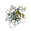
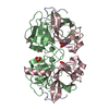
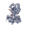
| Structure viewer | Molecule: MolmilJmol/JSmol |
|---|
- Downloads & links
-Download
| PDBx/mmCIF format | 2gct.cif.gz | 62.2 KB | Display | PDBx/mmCIF format |
|---|---|---|---|---|
| PDB format | pdb2gct.ent.gz | 45.1 KB | Display | PDB format |
| PDBx/mmJSON format | 2gct.json.gz | Tree view | PDBx/mmJSON format | |
| Others |  Other downloads Other downloads |
-Validation report
| Arichive directory | https://data.pdbj.org/pub/pdb/validation_reports/gc/2gctftp://data.pdbj.org/pub/pdb/validation_reports/gc/2gct | HTTPS FTP |
|---|
-Related structure data


-Links
 PDBj
PDBj

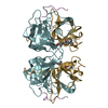
- Assembly
| Deposited unit | 
| ||||||||
|---|---|---|---|---|---|---|---|---|---|
| 1 | 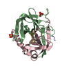
| ||||||||
| Unit cell |
|
-Components
-GAMMA-CHYMOTRYPSIN ... , 3 types, 3 molecules ABC
| #1: Protein/peptide | Mass: 1253.511 Da / Num. of mol.: 1 / Source method: isolated from a natural source / Source: (natural) |
|---|---|
| #2: Protein | Mass: 13934.556 Da / Num. of mol.: 1 / Source method: isolated from a natural source / Source: (natural) |
| #3: Protein | Mass: 10074.495 Da / Num. of mol.: 1 / Source method: isolated from a natural source / Source: (natural) |
-Protein/peptide , 1 types, 1 molecules D
| #4: Protein/peptide | Mass: 491.538 Da / Num. of mol.: 1 Source method: isolated from a genetically manipulated source |
|---|
-Non-polymers , 2 types, 133 molecules 

| #5: Chemical |  Mass: 96.063 Da / Num. of mol.: 2 / Source method: obtained synthetically / Formula: SO4 Mass: 96.063 Da / Num. of mol.: 2 / Source method: obtained synthetically / Formula: SO4#6: Water | ChemComp-HOH / | Mass: 18.015 Da / Num. of mol.: 131 / Source method: isolated from a natural source / Formula: H2O |
|---|
-Details
| Compound details | THE GAMMA CHYMOTRYPSIN MOLECULE IS COMPRISED OF THREE POLYPEPTIDE CHAINS WHICH ARE DERIVED FROM THE ...THE GAMMA CHYMOTRYPS |
|---|---|
| Has protein modification | Y |
-Experimental details
-Experiment
| Experiment | Method: X-RAY DIFFRACTION / Number of used crystals: 1 |
|---|
- Sample preparation
| Crystal | Density Matthews: 2.33 Å3/Da / Density % sol: 47.19 % |
|---|---|
| Crystal grow | pH: 2 / Details: pH 2.0 |
| Crystal grow | *PLUS pH: 7 / Method: batch method |
| Components of the solutions | *PLUS Conc.: 50 %sat / Common name: ammonium sulfate |
-Data collection
| Diffraction | Mean temperature: 285 K |
|---|---|
| Diffraction source | Source: ROTATING ANODE / Wavelength: 1.54 |
| Detector | Type: KODAK / Detector: FILM |
| Radiation | Protocol: SINGLE WAVELENGTH / Monochromatic (M) / Laue (L): M / Scattering type: x-ray |
| Radiation wavelength | Wavelength: 1.54 Å / Relative weight: 1 |
| Reflection | *PLUS Highest resolution: 1.8 Å / Lowest resolution: 10 Å / Num. obs: 13431 / % possible obs: 63.2 % / Num. measured all: 17029 / Rmerge(I) obs: 0.068 |
- Processing
| Software |
| ||||||||||||||||||||||||||||||||||||||||
|---|---|---|---|---|---|---|---|---|---|---|---|---|---|---|---|---|---|---|---|---|---|---|---|---|---|---|---|---|---|---|---|---|---|---|---|---|---|---|---|---|---|
| Refinement | Method to determine structure: MOLECULAR REPLACEMENT / Resolution: 1.8→10 Å / Rfactor Rwork: 0.169 | ||||||||||||||||||||||||||||||||||||||||
| Refinement step | Cycle: LAST / Resolution: 1.8→10 Å
| ||||||||||||||||||||||||||||||||||||||||
| Refine LS restraints |
| ||||||||||||||||||||||||||||||||||||||||
| Refine LS restraints | *PLUS
|
