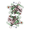
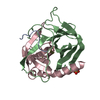
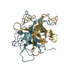
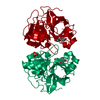
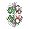
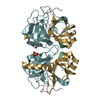
SHEET THE SHEET PRESENTED AS *S1* ON SHEET RECORDS BELOW IS ACTUALLY A SIX-STRANDED BETA-BARREL. ...SHEET THE SHEET PRESENTED AS *S1* ON SHEET RECORDS BELOW IS ACTUALLY A SIX-STRANDED BETA-BARREL. THIS IS REPRESENTED BY A SEVEN-STRANDED SHEET IN WHICH THE FIRST AND LAST STRANDS ARE IDENTICAL. SHEET S2 OF THIS MOLECULE IS BIFURCATED. IN ORDER TO REPRESENT THIS FEATURE IN THE SHEET RECORDS BELOW, TWO SHEETS ARE DEFINED. STRANDS 1, 2, 3, 4, 5, AND 7 OF SHEETS S2A AND S2B ARE IDENTICAL.
Mass: 18.015 Da / Num. of mol.: 141 / Source method: isolated from a natural source / Formula: H2O
-
Details
Compound details
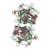
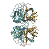
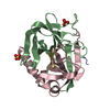
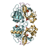
3GCT THE GAMMA CHYMOTRYPSIN MOLECULE IS COMPRISED OF THREE POLYPEPTIDE CHAINS WHICH ARE DERIVED ...3GCT THE GAMMA CHYMOTRYPSIN MOLECULE IS COMPRISED OF THREE POLYPEPTIDE CHAINS WHICH ARE DERIVED FROM THE ZYMOGEN OF THIS ENZYME BY EXCISION OF RESIDUES 14-15 AND 147-148. TO ASSIGN SEPARATE CHAIN IDENTIFIERS TO THE THREE CHAINS WOULD OBSCURE THIS RELATIONSHIP AND SO THIS WAS NOT DONE. CHAIN TERMINATOR RECORDS WERE INSERTED AFTER RESIDUES 146 AND 245 TO INDICATE EXPLICIT TERMINI AND THE SPECIAL CODE EXC WAS USED IN THE SEQRES RECORDS TO DENOTE THE EXCISIONS. RESIDUES 11 THROUGH 13 AND 149 THROUGH 150 ARE NOT VISIBLE IN THE ELECTRON DENSITY MAP AND SO ARE OMITTED. IN THE ABSENCE OF RESIDUE 13 THE TER RECORD WHICH WOULD HAVE APPEARED AFTER RESIDUE 13 IS ALSO OMITTED. RESIDUES B 500 - B 504 ARE A TETRAPEPTIDE BOUND IN THE ACTIVE SITE, COVALENTLY LINKED TO OG OF SER 195 AS AN ACYL ADDUCT. IT IS, PRESUMABLY, AN AUTOLYTIC CLEAVAGE PRODUCT. THE EXACT IDENTITY OF THE RESIDUES IS UNCERTAIN AS THE SIDE CHAINS SEEM TO BE DISORDERED. THE ATOM IDENTIFIED AS C UNK B 500 IS ACTUALLY THE CARBONYL CARBON OF AN UNIDENTIFIED AMINO ACID NOT VISIBLE IN THE ELECTRON DENSITY MAP.
Has protein modification
Y
-
Experimental details
-
Experiment
Experiment
Method: X-RAY DIFFRACTION / Number of used crystals: 1
-
Sample preparation
Crystal
Density Matthews: 2.32 Å3/Da / Density % sol: 47.08 %
Crystal grow
pH: 10.5 / Details: pH 10.5
Crystal grow
*PLUS
pH: 7 / Method: batch method
Components of the solutions
*PLUS
Conc.: 50 %sat / Common name: ammonium sulfate
-
Data collection
Diffraction
Mean temperature: 285 K
Diffraction source
Source: ROTATING ANODE / Wavelength: 1.54
Detector
Type: KODAK / Detector: FILM
Radiation
Protocol: SINGLE WAVELENGTH / Scattering type: x-ray
Radiation wavelength
Wavelength: 1.54 Å / Relative weight: 1
Reflection
*PLUS
Highest resolution: 1.6 Å / Lowest resolution: 10 Å / Num. obs: 23255 / % possible obs: 70.3 % / Num. measured all: 58476 / Rmerge(I) obs: 0.066
-
Processing
Software
Name
Classification
TNT
refinement
OSCTST
datareduction
AGROVATA/ROTAVATE
datascaling
Refinement
Method to determine structure: MOLECULAR REPLACEMENT / Resolution: 1.6→10 Å /
Rfactor
Num. reflection
Rwork
0.173
-
obs
-
22745
Refinement step
Cycle: LAST / Resolution: 1.6→10 Å
Protein
Nucleic acid
Ligand
Solvent
Total
Num. atoms
1767
0
5
141
1913
Refine LS restraints
Refine-ID
Type
Dev ideal
Weight
X-RAY DIFFRACTION
t_bond_d
0.2
X-RAY DIFFRACTION
t_angle_deg
3.1
X-RAY DIFFRACTION
t_dihedral_angle_d
17.3
0
X-RAY DIFFRACTION
t_incorr_chiral_ct
X-RAY DIFFRACTION
t_pseud_angle
0
X-RAY DIFFRACTION
t_trig_c_planes
0.021
X-RAY DIFFRACTION
t_gen_planes
0.02
X-RAY DIFFRACTION
t_it
0
X-RAY DIFFRACTION
t_nbd
Refine LS restraints
*PLUS
Refine-ID
Type
Dev ideal
X-RAY DIFFRACTION
t_bond_d
0.019
X-RAY DIFFRACTION
t_dihedral_angle_d
X-RAY DIFFRACTION
t_dihedral_angle_deg
17.5
X-RAY DIFFRACTION
t_plane_restr
0.022
+
About Yorodumi
-
News
-
Feb 9, 2022. New format data for meta-information of EMDB entries
New format data for meta-information of EMDB entries
Version 3 of the EMDB header file is now the official format.
The previous official version 1.9 will be removed from the archive.
In the structure databanks used in Yorodumi, some data are registered as the other names, "COVID-19 virus" and "2019-nCoV". Here are the details of the virus and the list of structure data.
Jan 31, 2019. EMDB accession codes are about to change! (news from PDBe EMDB page)
EMDB accession codes are about to change! (news from PDBe EMDB page)
The allocation of 4 digits for EMDB accession codes will soon come to an end. Whilst these codes will remain in use, new EMDB accession codes will include an additional digit and will expand incrementally as the available range of codes is exhausted. The current 4-digit format prefixed with “EMD-” (i.e. EMD-XXXX) will advance to a 5-digit format (i.e. EMD-XXXXX), and so on. It is currently estimated that the 4-digit codes will be depleted around Spring 2019, at which point the 5-digit format will come into force.
The EM Navigator/Yorodumi systems omit the EMD- prefix.
Related info.:Q: What is EMD? / ID/Accession-code notation in Yorodumi/EM Navigator
Yorodumi is a browser for structure data from EMDB, PDB, SASBDB, etc.
This page is also the successor to EM Navigator detail page, and also detail information page/front-end page for Omokage search.
The word "yorodu" (or yorozu) is an old Japanese word meaning "ten thousand". "mi" (miru) is to see.
Related info.:EMDB / PDB / SASBDB / Comparison of 3 databanks / Yorodumi Search / Aug 31, 2016. New EM Navigator & Yorodumi / Yorodumi Papers / Jmol/JSmol / Function and homology information / Changes in new EM Navigator and Yorodumi
 Movie
Movie Controller

 Yorodumi
 Open data
 Basic information
 Components
 Keywords
 Function and homology information
 X-RAY DIFFRACTION /
  Authors
 Citation
 Structure visualization
 Downloads & links
 Other downloads


 PDBj
PDBj

 Assembly
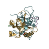


 Mass: 96.063 Da / Num. of mol.: 1 / Source method: obtained synthetically / Formula: SO4
 Sample preparation
 Processing