 Movie
Movie Controller

[English] 日本語
 Yorodumi
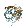
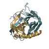
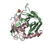
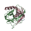
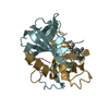
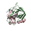
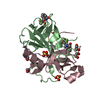
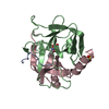
- PDB-1k2i: Crystal Structure of Gamma-Chymotrypsin in Complex with 7-Hydroxy... -
+ Open data

- Basic information
| Entry | Database: PDB / ID: 1k2i | ||||||
|---|---|---|---|---|---|---|---|
| Title | Crystal Structure of Gamma-Chymotrypsin in Complex with 7-Hydroxycoumarin | ||||||
 Components Components | CHYMOTRYPSINOGEN A | ||||||
 Keywords Keywords | HYDROLASE / enzyme-inhibitor complex | ||||||
| Function / homology |  Function and homology information Function and homology informationchymotrypsin / serpin family protein binding / serine protease inhibitor complex / digestion / serine-type endopeptidase activity / proteolysis / extracellular region Similarity search - Function | ||||||
| Biological species |  | ||||||
| Method |  X-RAY DIFFRACTION / FOURIER SYNTHESIS / Resolution: 1.8 Å X-RAY DIFFRACTION / FOURIER SYNTHESIS / Resolution: 1.8 Å | ||||||
 Authors Authors | Ghani, U. / Ng, K.K.S. / Atta-ur-Rahman / Choudhary, M.I. / Ullah, N. / James, M.N.G. | ||||||
 Citation Citation | Journal: J.Mol.Biol. / Year: 2001 Title: Crystal structure of gamma-chymotrypsin in complex with 7-hydroxycoumarin. Authors: Ghani, U. / Ng, K.K. / Atta-ur-Rahman / Choudhary, M.I. / Ullah, N. / James, M.N. | ||||||
| History |
|
- Structure visualization
| Structure viewer | Molecule: MolmilJmol/JSmol |
|---|
- Downloads & links
-Download
| PDBx/mmCIF format | 1k2i.cif.gz | 59.4 KB | Display | PDBx/mmCIF format |
|---|---|---|---|---|
| PDB format | pdb1k2i.ent.gz | 43 KB | Display | PDB format |
| PDBx/mmJSON format | 1k2i.json.gz | Tree view | PDBx/mmJSON format | |
| Others |  Other downloads Other downloads |
-Validation report
| Arichive directory | https://data.pdbj.org/pub/pdb/validation_reports/k2/1k2iftp://data.pdbj.org/pub/pdb/validation_reports/k2/1k2i | HTTPS FTP |
|---|
-Related structure data
| Related structure data |  1gctS S: Starting model for refinement |
|---|---|
| Similar structure data |


-Links
 PDBj
PDBj

- Assembly
| Deposited unit | 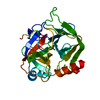
| ||||||||
|---|---|---|---|---|---|---|---|---|---|
| 1 |
| ||||||||
| 2 | 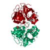
| ||||||||
| Unit cell |
| ||||||||
| Components on special symmetry positions |
|
-Components
| #1: Protein | Mass: 25686.037 Da / Num. of mol.: 1 / Source method: isolated from a natural source / Source: (natural) |
|---|---|
| #2: Chemical | ChemComp-SO4 /   Mass: 96.063 Da / Num. of mol.: 1 / Source method: obtained synthetically / Formula: SO4 Mass: 96.063 Da / Num. of mol.: 1 / Source method: obtained synthetically / Formula: SO4 |
| #3: Chemical | ChemComp-SN1 / 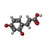  Mass: 180.157 Da / Num. of mol.: 1 / Source method: obtained synthetically / Formula: C9H8O4 Mass: 180.157 Da / Num. of mol.: 1 / Source method: obtained synthetically / Formula: C9H8O4 |
| #4: Water | ChemComp-HOH /  Mass: 18.015 Da / Num. of mol.: 134 / Source method: isolated from a natural source / Formula: H2O Mass: 18.015 Da / Num. of mol.: 134 / Source method: isolated from a natural source / Formula: H2O |
| Has protein modification | Y |
-Experimental details
-Experiment
| Experiment | Method: X-RAY DIFFRACTION / Number of used crystals: 1 |
|---|
- Sample preparation
| Crystal | Density Matthews: 2.31 Å3/Da / Density % sol: 46.79 % | ||||||||||||||||||||||||||||||
|---|---|---|---|---|---|---|---|---|---|---|---|---|---|---|---|---|---|---|---|---|---|---|---|---|---|---|---|---|---|---|---|
| Crystal grow | Temperature: 293 K / Method: vapor diffusion, hanging drop / pH: 7 Details: ammonium sulfate, sodium cacodylate, dioxane, pH 7.0, VAPOR DIFFUSION, HANGING DROP, temperature 293K | ||||||||||||||||||||||||||||||
| Crystal grow | *PLUS | ||||||||||||||||||||||||||||||
| Components of the solutions | *PLUS
|
-Data collection
| Diffraction | Mean temperature: 298 K |
|---|---|
| Diffraction source | Source: ROTATING ANODE / Type: RIGAKU RU200 / Wavelength: 1.5418 Å |
| Detector | Type: MACSCIENCE / Detector: IMAGE PLATE / Date: Aug 17, 1999 / Details: double-focusing mirrors |
| Radiation | Monochromator: Yale mirrors + Ni filter / Protocol: SINGLE WAVELENGTH / Monochromatic (M) / Laue (L): M / Scattering type: x-ray |
| Radiation wavelength | Wavelength: 1.5418 Å / Relative weight: 1 |
| Reflection | Resolution: 1.8→30 Å / Num. all: 23275 / Num. obs: 22972 / % possible obs: 99.7 % / Observed criterion σ(F): 0 / Observed criterion σ(I): -3 / Redundancy: 4.5 % / Biso Wilson estimate: 24.5 Å2 / Rmerge(I) obs: 0.084 / Rsym value: 0.084 / Net I/σ(I): 18.3 |
| Reflection shell | Resolution: 1.8→1.86 Å / Redundancy: 4.5 % / Rmerge(I) obs: 0.351 / Mean I/σ(I) obs: 4.3 / Num. unique all: 2229 / Rsym value: 0.351 / % possible all: 100 |
| Reflection | *PLUS Lowest resolution: 30 Å / Num. measured all: 104762 |
| Reflection shell | *PLUS Highest resolution: 1.8 Å / % possible obs: 100 % |
- Processing
| Software |
| |||||||||||||||||||||||||
|---|---|---|---|---|---|---|---|---|---|---|---|---|---|---|---|---|---|---|---|---|---|---|---|---|---|---|
| Refinement | Method to determine structure: FOURIER SYNTHESIS Starting model: PDB entry 1GCT Resolution: 1.8→30 Å / Isotropic thermal model: anisotropic / Cross valid method: THROUGHOUT / σ(F): 0 / σ(I): 0 / Stereochemistry target values: Engh & Huber
| |||||||||||||||||||||||||
| Displacement parameters | Biso mean: 16.7 Å2
| |||||||||||||||||||||||||
| Refinement step | Cycle: LAST / Resolution: 1.8→30 Å
| |||||||||||||||||||||||||
| Refine LS restraints |
| |||||||||||||||||||||||||
| Refinement | *PLUS Lowest resolution: 30 Å / Num. reflection obs: 22933 / Rfactor obs: 0.179 | |||||||||||||||||||||||||
| Solvent computation | *PLUS | |||||||||||||||||||||||||
| Displacement parameters | *PLUS |
