 Movie
Movie Controller

[English] 日本語
 Yorodumi
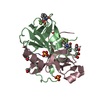
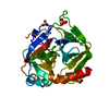
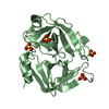
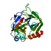
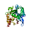
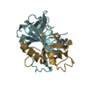
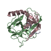
- PDB-1ggd: CRYSTAL STRUCTURE OF GAMMA CHYMOTRYPSIN WITH N-ACETYL-LEUCIL-PHEN... -
+ Open data

- Basic information
| Entry | Database: PDB / ID: 1ggd | ||||||
|---|---|---|---|---|---|---|---|
| Title | CRYSTAL STRUCTURE OF GAMMA CHYMOTRYPSIN WITH N-ACETYL-LEUCIL-PHENYLALANINE ALDEHYDE BOUND AT THE ACTIVE SITE | ||||||
 Components Components | (GAMMA CHYMOTRYPSIN) x 3 | ||||||
 Keywords Keywords | HYDROLASE/HYDROLASE INHIBITOR / CHYMOTRYPSIN / HYDROLASE-HYDROLASE INHIBITOR COMPLEX | ||||||
| Function / homology |  Function and homology information Function and homology informationchymotrypsin / serpin family protein binding / serine protease inhibitor complex / digestion / serine-type endopeptidase activity / proteolysis / extracellular region Similarity search - Function | ||||||
| Biological species |  | ||||||
| Method |  X-RAY DIFFRACTION / Resolution: 1.5 Å X-RAY DIFFRACTION / Resolution: 1.5 Å | ||||||
 Authors Authors | Neidhart, D. / Wei, Y. / Cassidy, C. / Lin, J. / Cleland, W.W. / Frey, P.A. | ||||||
 Citation Citation | Journal: Biochemistry / Year: 2000 Title: Correlation of low-barrier hydrogen bonding and oxyanion binding in transition state analogue complexes of chymotrypsin. Authors: Neidhart, D. / Wei, Y. / Cassidy, C. / Lin, J. / Cleland, W.W. / Frey, P.A. | ||||||
| History |
|
- Structure visualization
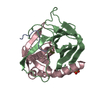
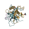
| Structure viewer | Molecule: MolmilJmol/JSmol |
|---|
- Downloads & links
-Download
| PDBx/mmCIF format | 1ggd.cif.gz | 63 KB | Display | PDBx/mmCIF format |
|---|---|---|---|---|
| PDB format | pdb1ggd.ent.gz | 45.1 KB | Display | PDB format |
| PDBx/mmJSON format | 1ggd.json.gz | Tree view | PDBx/mmJSON format | |
| Others |  Other downloads Other downloads |
-Validation report
| Arichive directory | https://data.pdbj.org/pub/pdb/validation_reports/gg/1ggdftp://data.pdbj.org/pub/pdb/validation_reports/gg/1ggd | HTTPS FTP |
|---|
-Related structure data


-Links
 PDBj
PDBj

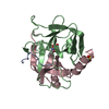
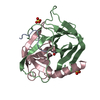
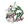
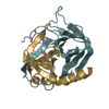
- Assembly
| Deposited unit | 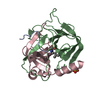
| ||||||||||||
|---|---|---|---|---|---|---|---|---|---|---|---|---|---|
| 1 |
| ||||||||||||
| 2 | 
| ||||||||||||
| 3 | 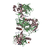
| ||||||||||||
| Unit cell |
| ||||||||||||
| Components on special symmetry positions |
|
-Components
-Protein/peptide , 1 types, 1 molecules A
| #1: Protein/peptide | Mass: 996.223 Da / Num. of mol.: 1 / Source method: isolated from a natural source / Source: (natural) |
|---|
-Protein , 2 types, 2 molecules BC
| #2: Protein | Mass: 13934.556 Da / Num. of mol.: 1 / Source method: isolated from a natural source / Source: (natural) |
|---|---|
| #3: Protein | Mass: 10074.495 Da / Num. of mol.: 1 / Source method: isolated from a natural source / Source: (natural) |
-Non-polymers , 3 types, 217 molecules 


| #4: Chemical | ChemComp-SO4 /  Mass: 96.063 Da / Num. of mol.: 1 / Source method: obtained synthetically / Formula: SO4 Mass: 96.063 Da / Num. of mol.: 1 / Source method: obtained synthetically / Formula: SO4 |
|---|---|
| #5: Chemical | ChemComp-FAF /  Mass: 304.384 Da / Num. of mol.: 1 / Source method: obtained synthetically / Formula: C17H24N2O3 Mass: 304.384 Da / Num. of mol.: 1 / Source method: obtained synthetically / Formula: C17H24N2O3 |
| #6: Water | ChemComp-HOH / Mass: 18.015 Da / Num. of mol.: 215 / Source method: isolated from a natural source / Formula: H2O |
-Details
| Has protein modification | Y |
|---|
-Experimental details
-Experiment
| Experiment | Method: X-RAY DIFFRACTION / Number of used crystals: 1 |
|---|
- Sample preparation
| Crystal | Density Matthews: 2.36 Å3/Da / Density % sol: 47.93 % | |||||||||||||||
|---|---|---|---|---|---|---|---|---|---|---|---|---|---|---|---|---|
| Crystal grow | Temperature: 298 K / Method: vapor diffusion / pH: 7 Details: ammonium sulfate, potassium phosphate, pH 7.0, vapor diffusion, temperature 298.0K | |||||||||||||||
| Crystal grow | *PLUS | |||||||||||||||
| Components of the solutions | *PLUS
|
-Data collection
| Diffraction | Mean temperature: 277 K |
|---|---|
| Diffraction source | Source: ROTATING ANODE / Type: RIGAKU RU200 / Wavelength: 1.5418 |
| Detector | Type: SIEMENS / Detector: AREA DETECTOR / Date: Nov 29, 1998 |
| Radiation | Protocol: SINGLE WAVELENGTH / Monochromatic (M) / Laue (L): M / Scattering type: x-ray |
| Radiation wavelength | Wavelength: 1.5418 Å / Relative weight: 1 |
| Reflection | Resolution: 1.5→30 Å / Num. all: 38043 / Num. obs: 34112 / % possible obs: 88 % / Observed criterion σ(I): 2 / Redundancy: 3.9 % / Rmerge(I) obs: 0.043 |
| Reflection shell | Resolution: 1.5→1.55 Å / Rmerge(I) obs: 0.184 / % possible all: 55.1 |
| Reflection | *PLUS Num. obs: 38043 |
| Reflection shell | *PLUS % possible obs: 55.1 % |
- Processing
| Software |
| |||||||||||||||||||||
|---|---|---|---|---|---|---|---|---|---|---|---|---|---|---|---|---|---|---|---|---|---|---|
| Refinement | Resolution: 1.5→8 Å / σ(F): 2 / Stereochemistry target values: Engh & Huber Details: Initial coordinates for this structure were derived from the model of G. Cohen et al. (PDB entry 2GCH). Secondary structure elements and sequence data are identical to those of entry 2GCH. ...Details: Initial coordinates for this structure were derived from the model of G. Cohen et al. (PDB entry 2GCH). Secondary structure elements and sequence data are identical to those of entry 2GCH. RESIDUES ALA 149 and ASN 150 ARE DISORDERED.
| |||||||||||||||||||||
| Refinement step | Cycle: LAST / Resolution: 1.5→8 Å
| |||||||||||||||||||||
| Refine LS restraints |
| |||||||||||||||||||||
| Software | *PLUS Name: X-PLOR / Version: 3.5 / Classification: refinement | |||||||||||||||||||||
| Refinement | *PLUS Highest resolution: 1.5 Å / Lowest resolution: 8 Å / σ(F): 2 / Rfactor obs: 0.182 | |||||||||||||||||||||
| Solvent computation | *PLUS | |||||||||||||||||||||
| Displacement parameters | *PLUS | |||||||||||||||||||||
| Refine LS restraints | *PLUS Type: x_angle_deg / Dev ideal: 1.3 |
