 Movie
Movie Controller

[English] 日本語
 Yorodumi
- PDB-2vgc: GAMMA-CHYMOTRYPSIN D-PARA-CHLORO-1-ACETAMIDO BORONIC ACID INHIBIT... -
+ Open data

- Basic information
| Entry | Database: PDB / ID: 2vgc | ||||||
|---|---|---|---|---|---|---|---|
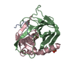
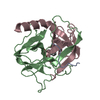
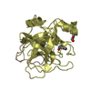
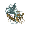
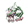
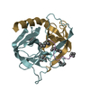
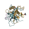
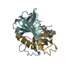
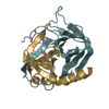
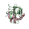
| Title | GAMMA-CHYMOTRYPSIN D-PARA-CHLORO-1-ACETAMIDO BORONIC ACID INHIBITOR COMPLEX | ||||||
 Components Components | (GAMMA CHYMOTRYPSIN) x 3 | ||||||
 Keywords Keywords | SERINE PROTEASE / HYDROLASE | ||||||
| Function / homology |  Function and homology information Function and homology informationchymotrypsin / serpin family protein binding / serine protease inhibitor complex / digestion / serine-type endopeptidase activity / proteolysis / extracellular region Similarity search - Function | ||||||
| Biological species |  | ||||||
| Method |  X-RAY DIFFRACTION / DIRECT SOLUTION WITH KNOWN STRUCTURE / Resolution: 1.8 Å X-RAY DIFFRACTION / DIRECT SOLUTION WITH KNOWN STRUCTURE / Resolution: 1.8 Å | ||||||
 Authors Authors | Stoll, V.S. / Eger, B.T. / Hynes, R.C. / Martichonok, V. / Jones, J.B. / Pai, E.F. | ||||||
 Citation Citation | Journal: Biochemistry / Year: 1998 Title: Differences in binding modes of enantiomers of 1-acetamido boronic acid based protease inhibitors: crystal structures of gamma-chymotrypsin and subtilisin Carlsberg complexes. Authors: Stoll, V.S. / Eger, B.T. / Hynes, R.C. / Martichonok, V. / Jones, J.B. / Pai, E.F. #1: Journal: J.Am.Chem.Soc. / Year: 1996Title: Probing the Specificity of the Serine Proteases Subtilisin Carlsberg and A-Chymotrypsin with Enantiomeric 1-Acetamido Boronic Acids. An Unexpected Reversal of the Normal Authors: Martichonok, V. / Jones, J.B. #2: Journal: Bioorg.Med.Chem. / Year: 1994Title: Probing the Specificity of the S1 Binding Site of Subtilisin Carlsberg with Boronic Acids Authors: Seufer-Wasserthal, P. / Martichonok, V. / Keller, T.H. / Chin, B. / Martin, R. / Jones, J.B. #3: Journal: Biochemistry / Year: 1991Title: Gamma-Chymotrypsin is a Complex of Alpha-Chymotrypsin with its Own Autolysis Products Authors: Harel, M. / Su, C.T. / Frolow, F. / Silman, I. / Sussman, J.L. #4: Journal: Biochemistry / Year: 1990Title: Structure and Activity of Two Photoreversible Cinnamates Bound to Chymotrypsin Authors: Stoddard, B.L. / Bruhnke, J. / Porter, N. / Ringe, D. / Petsko, G.A. | ||||||
| History |
|
- Structure visualization
| Structure viewer | Molecule: MolmilJmol/JSmol |
|---|
- Downloads & links
-Download
| PDBx/mmCIF format | 2vgc.cif.gz | 59.9 KB | Display | PDBx/mmCIF format |
|---|---|---|---|---|
| PDB format | pdb2vgc.ent.gz | 42.9 KB | Display | PDB format |
| PDBx/mmJSON format | 2vgc.json.gz | Tree view | PDBx/mmJSON format | |
| Others |  Other downloads Other downloads |
-Validation report
| Arichive directory | https://data.pdbj.org/pub/pdb/validation_reports/vg/2vgcftp://data.pdbj.org/pub/pdb/validation_reports/vg/2vgc | HTTPS FTP |
|---|
-Related structure data
| Related structure data | 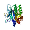 1av7C 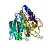 1avtC 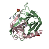 1vgcC 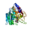 1vsbC 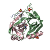 3vgcC 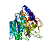 3vsbC 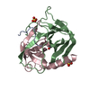 4vgcC 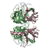 3gchS S: Starting model for refinement C: citing same article ( |
|---|---|
| Similar structure data |


-Links
 PDBj
PDBj

- Assembly
| Deposited unit | 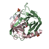
| ||||||||
|---|---|---|---|---|---|---|---|---|---|
| 1 |
| ||||||||
| 2 | 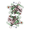
| ||||||||
| 3 | 
| ||||||||
| Unit cell |
|
-Components
-Protein/peptide , 1 types, 1 molecules A
| #1: Protein/peptide | Mass: 1253.511 Da / Num. of mol.: 1 / Source method: isolated from a natural source Details: A-CHYMOTRYPSIN PURCHASED FROM SIGMA AND CONVERTED TO G-CHYMOTRYPSIN BY THE METHOD OF STODDARD ET AL., 1990, BIOCHEMISTRY, VOL. 29, P. 4871-4879 Source: (natural) |
|---|
-Protein , 2 types, 2 molecules BC
| #2: Protein | Mass: 13934.556 Da / Num. of mol.: 1 / Source method: isolated from a natural source Details: A-CHYMOTRYPSIN PURCHASED FROM SIGMA AND CONVERTED TO G-CHYMOTRYPSIN BY THE METHOD OF STODDARD ET AL., 1990, BIOCHEMISTRY, VOL. 29, P. 4871-4879 Source: (natural) |
|---|---|
| #3: Protein | Mass: 10074.495 Da / Num. of mol.: 1 / Source method: isolated from a natural source Details: A-CHYMOTRYPSIN PURCHASED FROM SIGMA AND CONVERTED TO G-CHYMOTRYPSIN BY THE METHOD OF STODDARD ET AL., 1990, BIOCHEMISTRY, VOL. 29, P. 4871-4879 Source: (natural) |
-Non-polymers , 3 types, 98 molecules 

| #4: Chemical |  Mass: 96.063 Da / Num. of mol.: 2 / Source method: obtained synthetically / Formula: SO4 Mass: 96.063 Da / Num. of mol.: 2 / Source method: obtained synthetically / Formula: SO4#5: Chemical | ChemComp-V35 / |  Mass: 258.486 Da / Num. of mol.: 1 / Source method: obtained synthetically / Formula: C10H14BClNO4 Mass: 258.486 Da / Num. of mol.: 1 / Source method: obtained synthetically / Formula: C10H14BClNO4#6: Water | ChemComp-HOH / | Mass: 18.015 Da / Num. of mol.: 95 / Source method: isolated from a natural source / Formula: H2O |
|---|
-Details
| Has protein modification | Y |
|---|
-Experimental details
-Experiment
| Experiment | Method: X-RAY DIFFRACTION / Number of used crystals: 1 |
|---|
- Sample preparation
| Crystal | Density Matthews: 2.5 Å3/Da / Density % sol: 48 % | ||||||||||||||||||||||||||||||
|---|---|---|---|---|---|---|---|---|---|---|---|---|---|---|---|---|---|---|---|---|---|---|---|---|---|---|---|---|---|---|---|
| Crystal grow | pH: 6.5 Details: PROTEIN WAS CRYSTALLIZED IN SCINTILLATION VIALS IN 65% AMMONIUM SULFATE, 100 MM CACODYLATE, PH 6.5 | ||||||||||||||||||||||||||||||
| Crystal grow | *PLUS Details: Stoddard, B.L., (1990) Biochemistry, 29, 4871. | ||||||||||||||||||||||||||||||
| Components of the solutions | *PLUS
|
-Data collection
| Diffraction | Mean temperature: 287 K |
|---|---|
| Diffraction source | Source: ROTATING ANODE / Type: RIGAKU RUH2R / Wavelength: 1.5418 |
| Detector | Type: SIEMENS / Detector: AREA DETECTOR / Date: May 1, 1995 / Details: FRANKS MIRRORS |
| Radiation | Monochromator: NI FILTER / Monochromatic (M) / Laue (L): M / Scattering type: x-ray |
| Radiation wavelength | Wavelength: 1.5418 Å / Relative weight: 1 |
| Reflection | Resolution: 1.8→10 Å / Num. obs: 22354 / % possible obs: 79.69 % / Observed criterion σ(I): 1 / Redundancy: 3.47 % / Rsym value: 0.082 |
| Reflection shell | Resolution: 1.8→1.88 Å / Rsym value: 0.241 / % possible all: 56.49 |
| Reflection | *PLUS % possible obs: 88.4 % / Rmerge(I) obs: 0.082 |
- Processing
| Software |
| ||||||||||||||||||||||||||||||||||||||||||||||||||||||||||||
|---|---|---|---|---|---|---|---|---|---|---|---|---|---|---|---|---|---|---|---|---|---|---|---|---|---|---|---|---|---|---|---|---|---|---|---|---|---|---|---|---|---|---|---|---|---|---|---|---|---|---|---|---|---|---|---|---|---|---|---|---|---|
| Refinement | Method to determine structure: DIRECT SOLUTION WITH KNOWN STRUCTURE Starting model: PDB ENTRY 3GCH Resolution: 1.8→10 Å / Data cutoff high absF: 100000 / Data cutoff low absF: 0.1 / Cross valid method: THROUGHOUT / σ(F): 1
| ||||||||||||||||||||||||||||||||||||||||||||||||||||||||||||
| Refine analyze | Luzzati d res low obs: 10 Å | ||||||||||||||||||||||||||||||||||||||||||||||||||||||||||||
| Refinement step | Cycle: LAST / Resolution: 1.8→10 Å
| ||||||||||||||||||||||||||||||||||||||||||||||||||||||||||||
| Refine LS restraints |
| ||||||||||||||||||||||||||||||||||||||||||||||||||||||||||||
| LS refinement shell | Resolution: 1.8→1.88 Å / Total num. of bins used: 8
| ||||||||||||||||||||||||||||||||||||||||||||||||||||||||||||
| Xplor file |
| ||||||||||||||||||||||||||||||||||||||||||||||||||||||||||||
| Software | *PLUS Name: X-PLOR / Version: 3.1 / Classification: refinement | ||||||||||||||||||||||||||||||||||||||||||||||||||||||||||||
| Refinement | *PLUS Rfactor Rfree: 0.2542 | ||||||||||||||||||||||||||||||||||||||||||||||||||||||||||||
| Solvent computation | *PLUS | ||||||||||||||||||||||||||||||||||||||||||||||||||||||||||||
| Displacement parameters | *PLUS | ||||||||||||||||||||||||||||||||||||||||||||||||||||||||||||
| Refine LS restraints | *PLUS
|
