 Movie
Movie Controller

[English] 日本語
 Yorodumi
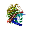
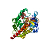
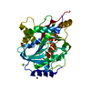
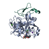
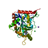
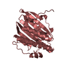
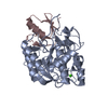
- PDB-1hja: LYS 18 VARIANT OF TURKEY OVOMUCOID INHIBITOR THIRD DOMAIN COMPLEX... -
+ Open data

- Basic information
| Entry | Database: PDB / ID: 1hja | ||||||
|---|---|---|---|---|---|---|---|
| Title | LYS 18 VARIANT OF TURKEY OVOMUCOID INHIBITOR THIRD DOMAIN COMPLEXED WITH ALPHA-CHYMOTRYPSIN | ||||||
 Components Components |
| ||||||
 Keywords Keywords | COMPLEX (HYDROLASE/INHIBITOR) / COMPLEX (HYDROLASE-INHIBITOR) / ALPHA-CHYMOTRYPSIN / PROTEIN INHIBITOR / COMPLEX (HYDROLASE-INHIBITOR) complex | ||||||
| Function / homology |  Function and homology information Function and homology informationchymotrypsin / molecular function inhibitor activity / serpin family protein binding / serine protease inhibitor complex / digestion / serine-type endopeptidase inhibitor activity / serine-type endopeptidase activity / proteolysis / extracellular region Similarity search - Function | ||||||
| Biological species |   | ||||||
| Method |  X-RAY DIFFRACTION / MOLECULAR REPLACEMENT / Resolution: 2.3 Å X-RAY DIFFRACTION / MOLECULAR REPLACEMENT / Resolution: 2.3 Å | ||||||
 Authors Authors | Ding, J.-H. / James, M.N.G. | ||||||
 Citation Citation | Journal: To be Published Title: Crystal Structure of Lys18 Variant of Turkey Ovomucoid Inhibitor Third Domain Complexed with Alpha-Chymotrypsin at 2.3 A Authors: Ding, J. / Qasim, M.A. / Laskowski Junior, M. / James, M.N.G. #1: Journal: J.Mol.Biol. / Year: 1987Title: Crystal and Molecular Structures of the Complex of Alpha-Chymotrypsin with its Inhibitor Turkey Ovomucoid Third Domain at 1.8 A Resolution Authors: Fujinaga, M. / Sielecki, A.R. / Read, R.J. / Ardelt, W. / Laskowski Junior, M. / James, M.N. | ||||||
| History |
|
- Structure visualization
| Structure viewer | Molecule: MolmilJmol/JSmol |
|---|
- Downloads & links
-Download
| PDBx/mmCIF format | 1hja.cif.gz | 68.6 KB | Display | PDBx/mmCIF format |
|---|---|---|---|---|
| PDB format | pdb1hja.ent.gz | 50 KB | Display | PDB format |
| PDBx/mmJSON format | 1hja.json.gz | Tree view | PDBx/mmJSON format | |
| Others |  Other downloads Other downloads |
-Validation report
| Arichive directory | https://data.pdbj.org/pub/pdb/validation_reports/hj/1hjaftp://data.pdbj.org/pub/pdb/validation_reports/hj/1hja | HTTPS FTP |
|---|
-Related structure data
| Related structure data | 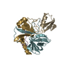 1choS S: Starting model for refinement |
|---|---|
| Similar structure data |


-Links
 PDBj
PDBj

- Assembly
| Deposited unit | 
| ||||||||
|---|---|---|---|---|---|---|---|---|---|
| 1 |
| ||||||||
| Unit cell |
|
-Components
| #1: Protein/peptide | Mass: 1253.511 Da / Num. of mol.: 1 / Source method: isolated from a natural source Details: COMMERCIAL PRODUCT OF WORTHINGTON BIOCHEMICAL CORPORATION Source: (natural) |
|---|---|
| #2: Protein | Mass: 13934.556 Da / Num. of mol.: 1 Source method: isolated from a genetically manipulated source Source: (gene. exp.) Description: COMMERCIAL PRODUCT OF WORTHINGTON BIOCHEMICAL CORPORATION Plasmid: PEZZ318.TKY / Production host:  |
| #3: Protein | Mass: 10074.495 Da / Num. of mol.: 1 Source method: isolated from a genetically manipulated source Source: (gene. exp.) Description: COMMERCIAL PRODUCT OF WORTHINGTON BIOCHEMICAL CORPORATION Plasmid: PEZZ318.TKY / Production host: |
| #4: Protein | Mass: 5601.311 Da / Num. of mol.: 1 Fragment: THIRD DOMAIN, DELETION OF FIRST 5 RESIDUES FROM N-TERMINUS Mutation: DEL(1-5), L18K Source method: isolated from a genetically manipulated source Details: TURKEY OVOMUCOID INHIBITOR / Source: (gene. exp.) |
| #5: Water | ChemComp-HOH /  Mass: 18.015 Da / Num. of mol.: 99 / Source method: isolated from a natural source / Formula: H2O Mass: 18.015 Da / Num. of mol.: 99 / Source method: isolated from a natural source / Formula: H2O |
| Has protein modification | Y |
-Experimental details
-Experiment
| Experiment | Method: X-RAY DIFFRACTION / Number of used crystals: 1 |
|---|
- Sample preparation
| Crystal | Density Matthews: 2.48 Å3/Da / Density % sol: 47.84 % Description: ALPHA-CHYMOTRYPSIN (E.C.3.4.21.1) COMPLEX WITH TURKEY OVOMUCOID THIRD DOMAIN |
|---|---|
| Crystal grow | pH: 6 / Details: 0.1 M KH2PO4/K2HPO4 PH=6.0 10% PEG6000 |
-Data collection
| Diffraction | Mean temperature: 293 K |
|---|---|
| Diffraction source | Source: ROTATING ANODE / Type: OTHER / Wavelength: 1.5418 |
| Detector | Type: SIEMENS / Detector: AREA DETECTOR / Date: Nov 4, 1996 |
| Radiation | Monochromator: GRAPHITE(002) / Monochromatic (M) / Laue (L): M / Scattering type: x-ray |
| Radiation wavelength | Wavelength: 1.5418 Å / Relative weight: 1 |
| Reflection | Resolution: 2.3→10 Å / Num. obs: 11136 / % possible obs: 86.9 % / Observed criterion σ(I): 1 / Redundancy: 8.7 % / Biso Wilson estimate: 32.2 Å2 / Rmerge(I) obs: 0.108 / Net I/σ(I): 5.89 |
| Reflection shell | Resolution: 2.3→2.4 Å / % possible all: 69.27 |
- Processing
| Software |
| ||||||||||||||||||||||||||||||||||||||||||||||||||||||||||||
|---|---|---|---|---|---|---|---|---|---|---|---|---|---|---|---|---|---|---|---|---|---|---|---|---|---|---|---|---|---|---|---|---|---|---|---|---|---|---|---|---|---|---|---|---|---|---|---|---|---|---|---|---|---|---|---|---|---|---|---|---|---|
| Refinement | Method to determine structure: MOLECULAR REPLACEMENT Starting model: PDB ENTRY 1CHO Resolution: 2.3→10 Å / σ(F): 1
| ||||||||||||||||||||||||||||||||||||||||||||||||||||||||||||
| Displacement parameters | Biso mean: 14.55 Å2 | ||||||||||||||||||||||||||||||||||||||||||||||||||||||||||||
| Refine analyze | Luzzati sigma a obs: 0.36 Å | ||||||||||||||||||||||||||||||||||||||||||||||||||||||||||||
| Refinement step | Cycle: LAST / Resolution: 2.3→10 Å
| ||||||||||||||||||||||||||||||||||||||||||||||||||||||||||||
| Refine LS restraints |
| ||||||||||||||||||||||||||||||||||||||||||||||||||||||||||||
| LS refinement shell | Resolution: 2.3→2.4 Å / Total num. of bins used: 8
|
