Protocol: SINGLE WAVELENGTH / Monochromatic (M) / Laue (L): M / Scattering type: x-ray
Radiation wavelength
Wavelength: 0.919 Å / Relative weight: 1
Reflection
Resolution: 1.7→20 Å / Num. obs: 39479 / % possible obs: 93.1 % / Redundancy: 4.1 % / Biso Wilson estimate: 16 Å2 / Rsym value: 0.045 / Net I/σ(I): 12.4
Reflection shell
Resolution: 1.7→1.79 Å / Redundancy: 2.2 % / Mean I/σ(I) obs: 8.1 / Rsym value: 0.078 / % possible all: 83.7
Reflection
*PLUS
Num. measured all: 276082 / Rmerge(I) obs: 0.045
Reflection shell
*PLUS
% possible obs: 83.7 % / Rmerge(I) obs: 0.078
-
Processing
Software
Name
Version
Classification
MADSYS
phasing
SHELX
modelbuilding
MLPHARE
phasing
X-PLOR
3.843
refinement
MOSFLM
datareduction
CCP4
(SCALA)
datascaling
SHELX
phasing
Refinement
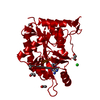
Method to determine structure: MAD / Resolution: 1.7→20 Å / Rfactor Rfree error: 0.003 / Data cutoff high absF: 1000000 / Data cutoff low absF: 0.001 / Cross valid method: THROUGHOUT / σ(F): 2 Details: RESIDUES 282 AND 283 WERE NOT SEEN IN THE ELECTRON DENSITY MAPS DUE TO DISORDER
Rfactor
Num. reflection
% reflection
Selection details
Rfree
0.202
3806
10 %
RANDOM
Rwork
0.181
-
-
-
obs
-
38017
92.8 %
-
Displacement parameters
Biso mean: 17.3 Å2
Refine analyze
Free
Obs
Luzzati coordinate error
0.2 Å
0.17 Å
Luzzati d res low
-
5 Å
Refinement step
Cycle: LAST / Resolution: 1.7→20 Å
Protein
Nucleic acid
Ligand
Solvent
Total
Num. atoms
2066
0
10
173
2249
Refine LS restraints
Refine-ID
Type
Dev ideal
X-RAY DIFFRACTION
x_bond_d
0.007
X-RAY DIFFRACTION
x_bond_d_na
X-RAY DIFFRACTION
x_bond_d_prot
X-RAY DIFFRACTION
x_angle_d
X-RAY DIFFRACTION
x_angle_d_na
X-RAY DIFFRACTION
x_angle_d_prot
X-RAY DIFFRACTION
x_angle_deg
1.258
X-RAY DIFFRACTION
x_angle_deg_na
X-RAY DIFFRACTION
x_angle_deg_prot
X-RAY DIFFRACTION
x_dihedral_angle_d
23.8
X-RAY DIFFRACTION
x_dihedral_angle_d_na
X-RAY DIFFRACTION
x_dihedral_angle_d_prot
X-RAY DIFFRACTION
x_improper_angle_d
1.18
X-RAY DIFFRACTION
x_improper_angle_d_na
X-RAY DIFFRACTION
x_improper_angle_d_prot
X-RAY DIFFRACTION
x_mcbond_it
X-RAY DIFFRACTION
x_mcangle_it
X-RAY DIFFRACTION
x_scbond_it
X-RAY DIFFRACTION
x_scangle_it
LS refinement shell
Resolution: 1.7→1.78 Å / Rfactor Rfree error: 0.013 / Total num. of bins used: 8
In the structure databanks used in Yorodumi, some data are registered as the other names, "COVID-19 virus" and "2019-nCoV". Here are the details of the virus and the list of structure data.
Jan 31, 2019. EMDB accession codes are about to change! (news from PDBe EMDB page)
EMDB accession codes are about to change! (news from PDBe EMDB page)
The allocation of 4 digits for EMDB accession codes will soon come to an end. Whilst these codes will remain in use, new EMDB accession codes will include an additional digit and will expand incrementally as the available range of codes is exhausted. The current 4-digit format prefixed with “EMD-” (i.e. EMD-XXXX) will advance to a 5-digit format (i.e. EMD-XXXXX), and so on. It is currently estimated that the 4-digit codes will be depleted around Spring 2019, at which point the 5-digit format will come into force.
The EM Navigator/Yorodumi systems omit the EMD- prefix.
Related info.:Q: What is EMD? / ID/Accession-code notation in Yorodumi/EM Navigator
Yorodumi is a browser for structure data from EMDB, PDB, SASBDB, etc.
This page is also the successor to EM Navigator detail page, and also detail information page/front-end page for Omokage search.
The word "yorodu" (or yorozu) is an old Japanese word meaning "ten thousand". "mi" (miru) is to see.
Related info.:EMDB / PDB / SASBDB / Comparison of 3 databanks / Yorodumi Search / Aug 31, 2016. New EM Navigator & Yorodumi / Yorodumi Papers / Jmol/JSmol / Function and homology information / Changes in new EM Navigator and Yorodumi
 Movie
Movie Controller

 Yorodumi
 Open data
 Basic information
 Components
 Keywords
 Function and homology information
 Homo sapiens (human)
 X-RAY DIFFRACTION /
  Authors
 Citation
 Structure visualization
 Downloads & links
 Other downloads
 PDBj
PDBj


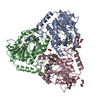
 Assembly


 Mass: 135.127 Da / Num. of mol.: 1 / Source method: obtained synthetically / Formula: C5H5N5
 Mass: 18.015 Da / Num. of mol.: 173 / Source method: isolated from a natural source / Formula: H2O
 Sample preparation
 / Beamline: F1 / Wavelength: 0.919
  Processing