 Movie
Movie Controller

[English] 日本語
 Yorodumi
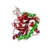
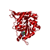
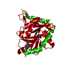
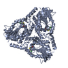
- PDB-1k27: Crystal Structure of 5'-Deoxy-5'-Methylthioadenosine Phosphorylas... -
+ Open data

- Basic information
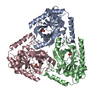
| Entry | Database: PDB / ID: 1k27 | ||||||
|---|---|---|---|---|---|---|---|
| Title | Crystal Structure of 5'-Deoxy-5'-Methylthioadenosine Phosphorylase in Complex with a Transition State Analogue | ||||||
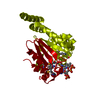
 Components Components | 5'-Deoxy-5'-Methylthioadenosine Phosphorylase | ||||||
 Keywords Keywords | TRANSFERASE / MTAP / methylthioadenosine phosphorylase / transition state analogue / phosphate | ||||||
| Function / homology |  Function and homology information Function and homology informationMethionine salvage pathway / 1,4-alpha-oligoglucan phosphorylase activity / S-methyl-5'-thioadenosine phosphorylase / S-methyl-5-thioadenosine phosphorylase activity / : / purine ribonucleoside salvage / nucleobase-containing compound metabolic process / Gene and protein expression by JAK-STAT signaling after Interleukin-12 stimulation / extracellular exosome / nucleus / cytosol Similarity search - Function | ||||||
| Biological species |  Homo sapiens (human) Homo sapiens (human) | ||||||
| Method |  X-RAY DIFFRACTION / SYNCHROTRON / FOURIER SYNTHESIS / Resolution: 1.95 Å X-RAY DIFFRACTION / SYNCHROTRON / FOURIER SYNTHESIS / Resolution: 1.95 Å | ||||||
 Authors Authors | Shi, W. / Singh, V. / Tyler, P.C. / Furneaux, R.H. / Almo, S.C. / Schramm, V.L. | ||||||
 Citation Citation | Journal: Biochemistry / Year: 2004 Title: Picomolar transition state analogue inhibitors of human 5'-methylthioadenosine phosphorylase and X-ray structure with MT-immucillin-A Authors: Singh, V. / Shi, W. / Evans, G.B. / Tyler, P.C. / Furneaux, R.H. / Almo, S.C. / Schramm, V.L. | ||||||
| History |
|
- Structure visualization
| Structure viewer | Molecule: MolmilJmol/JSmol |
|---|
- Downloads & links
-Download
| PDBx/mmCIF format | 1k27.cif.gz | 69.2 KB | Display | PDBx/mmCIF format |
|---|---|---|---|---|
| PDB format | pdb1k27.ent.gz | 50.1 KB | Display | PDB format |
| PDBx/mmJSON format | 1k27.json.gz | Tree view | PDBx/mmJSON format | |
| Others |  Other downloads Other downloads |
-Validation report
| Arichive directory | https://data.pdbj.org/pub/pdb/validation_reports/k2/1k27ftp://data.pdbj.org/pub/pdb/validation_reports/k2/1k27 | HTTPS FTP |
|---|
-Related structure data
| Related structure data |  1cg6S S: Starting model for refinement |
|---|---|
| Similar structure data |

-Links
 PDBj
PDBj


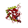
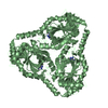
- Assembly
| Deposited unit | 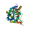
| ||||||||
|---|---|---|---|---|---|---|---|---|---|
| 1 | 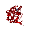
| ||||||||
| Unit cell |
| ||||||||
| Components on special symmetry positions |
| ||||||||
| Details | The other two monomers of the biological assembly is generated by the three fold axis, -Y, X-Y, Z; and -X+Y, -X, Z. |
-Components
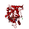
| #1: Protein | Mass: 31277.053 Da / Num. of mol.: 1 Source method: isolated from a genetically manipulated source Source: (gene. exp.) Homo sapiens (human) / Plasmid: PET-28A / Species (production host): Escherichia coli / Production host:  References: UniProt: Q13126, S-methyl-5'-thioadenosine phosphorylase |
|---|---|
| #2: Chemical | ChemComp-PO4 /   Mass: 94.971 Da / Num. of mol.: 1 / Source method: obtained synthetically / Formula: PO4 Mass: 94.971 Da / Num. of mol.: 1 / Source method: obtained synthetically / Formula: PO4 |
| #3: Chemical | ChemComp-MTM / (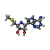  Mass: 297.377 Da / Num. of mol.: 1 / Source method: obtained synthetically / Formula: C12H19N5O2S Mass: 297.377 Da / Num. of mol.: 1 / Source method: obtained synthetically / Formula: C12H19N5O2S |
| #4: Water | ChemComp-HOH /  Mass: 18.015 Da / Num. of mol.: 138 / Source method: isolated from a natural source / Formula: H2O Mass: 18.015 Da / Num. of mol.: 138 / Source method: isolated from a natural source / Formula: H2O |
-Experimental details
-Experiment
| Experiment | Method: X-RAY DIFFRACTION / Number of used crystals: 1 |
|---|
- Sample preparation
| Crystal | Density Matthews: 3.08 Å3/Da / Density % sol: 60.13 % | ||||||||||||||||||||||||||||||
|---|---|---|---|---|---|---|---|---|---|---|---|---|---|---|---|---|---|---|---|---|---|---|---|---|---|---|---|---|---|---|---|
| Crystal grow | Temperature: 298 K / Method: vapor diffusion, hanging drop / pH: 8 Details: PEG 6000, Spermidine, Tris, pH 8.0, VAPOR DIFFUSION, HANGING DROP, temperature 298K | ||||||||||||||||||||||||||||||
| Crystal grow | *PLUS Temperature: 18 ℃ / Method: vapor diffusion, hanging drop | ||||||||||||||||||||||||||||||
| Components of the solutions | *PLUS
|
-Data collection
| Diffraction | Mean temperature: 100 K |
|---|---|
| Diffraction source | Source: SYNCHROTRON / Site: NSLS  / Beamline: X9B / Wavelength: 0.98 Å / Beamline: X9B / Wavelength: 0.98 Å |
| Detector | Type: ADSC QUANTUM 4 / Detector: CCD / Date: Mar 27, 2001 |
| Radiation | Protocol: SINGLE WAVELENGTH / Monochromatic (M) / Laue (L): M / Scattering type: x-ray |
| Radiation wavelength | Wavelength: 0.98 Å / Relative weight: 1 |
| Reflection | Resolution: 1.95→20 Å / Num. all: 28021 / Num. obs: 28021 / % possible obs: 99.5 % / Observed criterion σ(F): 0 / Observed criterion σ(I): 0 / Redundancy: 6 % / Biso Wilson estimate: 7.7 Å2 / Rsym value: 0.064 / Net I/σ(I): 14.6 |
| Reflection shell | Resolution: 1.95→2.02 Å / Redundancy: 5.8 % / Mean I/σ(I) obs: 3.6 / Num. unique all: 2794 / Rsym value: 0.387 / % possible all: 100 |
| Reflection | *PLUS Lowest resolution: 20 Å / Num. measured all: 167189 / Rmerge(I) obs: 0.064 |
| Reflection shell | *PLUS % possible obs: 100 % / Rmerge(I) obs: 0.386 |
- Processing
| Software |
| ||||||||||||||||||||||||||||||||||||
|---|---|---|---|---|---|---|---|---|---|---|---|---|---|---|---|---|---|---|---|---|---|---|---|---|---|---|---|---|---|---|---|---|---|---|---|---|---|
| Refinement | Method to determine structure: FOURIER SYNTHESIS Starting model: 1CG6 Resolution: 1.95→20 Å / Isotropic thermal model: Restrained / Cross valid method: THROUGHOUT / σ(F): 2 / Stereochemistry target values: Engh & Huber
| ||||||||||||||||||||||||||||||||||||
| Solvent computation | Solvent model: flat model / Bsol: 44.3308 Å2 / ksol: 0.406563 e/Å3 | ||||||||||||||||||||||||||||||||||||
| Displacement parameters | Biso mean: 19.9 Å2
| ||||||||||||||||||||||||||||||||||||
| Refine analyze |
| ||||||||||||||||||||||||||||||||||||
| Refinement step | Cycle: LAST / Resolution: 1.95→20 Å
| ||||||||||||||||||||||||||||||||||||
| Refine LS restraints |
| ||||||||||||||||||||||||||||||||||||
| LS refinement shell | Resolution: 1.95→2.07 Å / Rfactor Rfree error: 0.011 / Total num. of bins used: 6
| ||||||||||||||||||||||||||||||||||||
| Refinement | *PLUS Lowest resolution: 20 Å / Rfactor Rwork: 0.18 | ||||||||||||||||||||||||||||||||||||
| Solvent computation | *PLUS | ||||||||||||||||||||||||||||||||||||
| Displacement parameters | *PLUS | ||||||||||||||||||||||||||||||||||||
| Refine LS restraints | *PLUS
|
