 Movie
Movie Controller

[English] 日本語
 Yorodumi
- PDB-2cga: BOVINE CHYMOTRYPSINOGEN A. X-RAY CRYSTAL STRUCTURE ANALYSIS AND R... -
+ Open data

- Basic information
| Entry | Database: PDB / ID: 2cga | ||||||
|---|---|---|---|---|---|---|---|
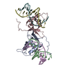
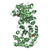
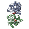
| Title | BOVINE CHYMOTRYPSINOGEN A. X-RAY CRYSTAL STRUCTURE ANALYSIS AND REFINEMENT OF A NEW CRYSTAL FORM AT 1.8 ANGSTROMS RESOLUTION | ||||||
 Components Components | CHYMOTRYPSINOGEN A | ||||||
 Keywords Keywords | HYDROLASE(ZYMOGEN) | ||||||
| Function / homology |  Function and homology information Function and homology informationchymotrypsin / serpin family protein binding / serine protease inhibitor complex / digestion / serine-type endopeptidase activity / proteolysis / extracellular region Similarity search - Function | ||||||
| Biological species |  | ||||||
| Method |  X-RAY DIFFRACTION / Resolution: 1.8 Å X-RAY DIFFRACTION / Resolution: 1.8 Å | ||||||
 Authors Authors | Wang, D. / Bode, W. / Huber, R. | ||||||
 Citation Citation | Journal: J.Mol.Biol. / Year: 1985 Title: Bovine chymotrypsinogen A X-ray crystal structure analysis and refinement of a new crystal form at 1.8 A resolution. Authors: Wang, D. / Bode, W. / Huber, R. #1: Journal: J.Mol.Biol. / Year: 1979Title: The Transition of Bovine Trypsinogen to a Trypsin-Like State Upon Strong Ligand Binding. II. The Binding of the Pancreatic Trypsin Inhibitor and of Isoleucine-Valine and of Sequentially ...Title: The Transition of Bovine Trypsinogen to a Trypsin-Like State Upon Strong Ligand Binding. II. The Binding of the Pancreatic Trypsin Inhibitor and of Isoleucine-Valine and of Sequentially Related Peptides to Trypsinogen and to P-Guanidinobenzoate-Trypsinogen Authors: Bode, W. #2: Journal: J.Mol.Biol. / Year: 1977Title: Crystal Structure of Bovine Trypsinogen at 1.8 Angstroms Resolution. II. Crystallographic Refinement, Refined Crystal Structure and Comparison with Bovine Trypsin Authors: Fehlhammer, H. / Bode, W. / Huber, R. #3: Journal: J.Mol.Biol. / Year: 1976Title: Crystal Structure of Bovine Trypsinogen at 1.8 Angstroms Resolution. I. Data Collection, Application of Patterson Search Techniques and Preliminary Structural Interpretation Authors: Bode, W. / Fehlhammer, H. / Huber, R. #4: Journal: Biochemistry / Year: 1970Title: Chymotrypsinogen. 2.5-Angstroms Crystal Structure, Comparison with Alpha-Chymotrypsin, and Implications for Zymogen Activation Authors: Freer, S.T. / Kraut, J. / Robertus, J.D. / Wright, H.T. / Xuong, N.H. | ||||||
| History |
|
- Structure visualization
| Structure viewer | Molecule: MolmilJmol/JSmol |
|---|
- Downloads & links
-Download
| PDBx/mmCIF format | 2cga.cif.gz | 99.3 KB | Display | PDBx/mmCIF format |
|---|---|---|---|---|
| PDB format | pdb2cga.ent.gz | 78.6 KB | Display | PDB format |
| PDBx/mmJSON format | 2cga.json.gz | Tree view | PDBx/mmJSON format | |
| Others |  Other downloads Other downloads |
-Validation report
| Arichive directory | https://data.pdbj.org/pub/pdb/validation_reports/cg/2cgaftp://data.pdbj.org/pub/pdb/validation_reports/cg/2cga | HTTPS FTP |
|---|
-Related structure data
| Similar structure data |
|---|


-Links
 PDBj
PDBj

- Assembly
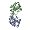
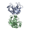
| Deposited unit | 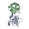
| ||||||||
|---|---|---|---|---|---|---|---|---|---|
| 1 |
| ||||||||
| Unit cell |
| ||||||||
| Atom site foot note | 1: RESIDUES VAL A 17 AND VAL B 17 HAVE UNUSUAL MAIN CHAIN CONFORMATION. | ||||||||
| Noncrystallographic symmetry (NCS) | NCS oper: (Code: given Matrix: (0.9877, 0.155, 0.0177), Vector: Details | THE TRANSFORMATION PRESENTED ON THE *MTRIX* RECORDS BELOW WILL YIELD COORDINATES FOR CHAIN *A* WHEN APPLIED TO CHAIN *B*. | |
-Components
| #1: Protein | Mass: 25686.037 Da / Num. of mol.: 2 Source method: isolated from a genetically manipulated source Source: (gene. exp.) #2: Water | ChemComp-HOH / |  Mass: 18.015 Da / Num. of mol.: 329 / Source method: isolated from a natural source / Formula: H2O Mass: 18.015 Da / Num. of mol.: 329 / Source method: isolated from a natural source / Formula: H2OHas protein modification | Y | |
|---|
-Experimental details
-Experiment
| Experiment | Method: X-RAY DIFFRACTION |
|---|
- Sample preparation
| Crystal | Density Matthews: 2.45 Å3/Da / Density % sol: 49.77 % | ||||||||||||||||||||
|---|---|---|---|---|---|---|---|---|---|---|---|---|---|---|---|---|---|---|---|---|---|
| Crystal grow | *PLUS pH: 4.5 / Method: vapor diffusion | ||||||||||||||||||||
| Components of the solutions | *PLUS
|
-Data collection
| Reflection | *PLUS Highest resolution: 1.8 Å / Num. obs: 32818 / % possible obs: 69 % / Observed criterion σ(I): 2 / Rmerge(I) obs: 0.086 / Num. measured all: 87718 |
|---|
- Processing
| Software | Name: EREF / Classification: refinement | ||||||||||||||||||||||||||||||||||||||||||||||||||||||||||||
|---|---|---|---|---|---|---|---|---|---|---|---|---|---|---|---|---|---|---|---|---|---|---|---|---|---|---|---|---|---|---|---|---|---|---|---|---|---|---|---|---|---|---|---|---|---|---|---|---|---|---|---|---|---|---|---|---|---|---|---|---|---|
| Refinement | Resolution: 1.8→6 Å /
| ||||||||||||||||||||||||||||||||||||||||||||||||||||||||||||
| Refinement step | Cycle: LAST / Resolution: 1.8→6 Å
| ||||||||||||||||||||||||||||||||||||||||||||||||||||||||||||
| Refine LS restraints |
| ||||||||||||||||||||||||||||||||||||||||||||||||||||||||||||
| Refinement | *PLUS Highest resolution: 1.8 Å / Lowest resolution: 6 Å / Num. reflection obs: 30617 / Rfactor obs: 0.173 | ||||||||||||||||||||||||||||||||||||||||||||||||||||||||||||
| Solvent computation | *PLUS | ||||||||||||||||||||||||||||||||||||||||||||||||||||||||||||
| Displacement parameters | *PLUS | ||||||||||||||||||||||||||||||||||||||||||||||||||||||||||||
| Refine LS restraints | *PLUS Type: o_angle_d |
