 Movie
Movie Controller

[English] 日本語
 Yorodumi
- PDB-1t8o: CRYSTAL STRUCTURE OF THE P1 TRP BPTI MUTANT- BOVINE CHYMOTRYPSIN ... -
+ Open data

- Basic information
| Entry | Database: PDB / ID: 1t8o | ||||||
|---|---|---|---|---|---|---|---|
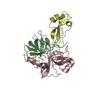
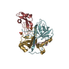
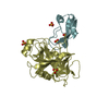
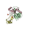
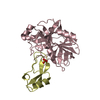
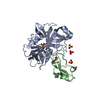
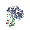
| Title | CRYSTAL STRUCTURE OF THE P1 TRP BPTI MUTANT- BOVINE CHYMOTRYPSIN COMPLEX | ||||||
 Components Components |
| ||||||
 Keywords Keywords | HYDROLASE/HYDROLASE INHIBITOR / CHYMOTRYPSIN / SERINE PROTEINASE / BOVINE PANCREATIC TRYPSIN INHIBITOR / BPTI / PROTEIN-PROTEIN INTERACTION / NON-COGNATE BINDING / S1 POCKET / PRIMARY SPECIFICITY / HYDROLASE-HYDROLASE INHIBITOR COMPLEX | ||||||
| Function / homology |  Function and homology information Function and homology informationchymotrypsin / sulfate binding / negative regulation of platelet aggregation / zymogen binding / potassium channel inhibitor activity / molecular function inhibitor activity / negative regulation of thrombin-activated receptor signaling pathway / serpin family protein binding / serine protease inhibitor complex / digestion ...chymotrypsin / sulfate binding / negative regulation of platelet aggregation / zymogen binding / potassium channel inhibitor activity / molecular function inhibitor activity / negative regulation of thrombin-activated receptor signaling pathway / serpin family protein binding / serine protease inhibitor complex / digestion / serine-type endopeptidase inhibitor activity / protease binding / serine-type endopeptidase activity / calcium ion binding / proteolysis / : / extracellular region Similarity search - Function | ||||||
| Biological species |  | ||||||
| Method |  X-RAY DIFFRACTION / SYNCHROTRON / MOLECULAR REPLACEMENT / Resolution: 1.7 Å X-RAY DIFFRACTION / SYNCHROTRON / MOLECULAR REPLACEMENT / Resolution: 1.7 Å | ||||||
 Authors Authors | Czapinska, H. / Helland, R. / Otlewski, J. / Smalas, A.O. | ||||||
 Citation Citation | Journal: J.Mol.Biol. / Year: 2004 Title: Crystal structures of five bovine chymotrypsin complexes with P1 BPTI variants. Authors: Czapinska, H. / Helland, R. / Smalas, A.O. / Otlewski, J. #1: Journal: J.Mol.Biol. / Year: 2003Title: STRUCTURAL CONSEQUENCES OF ACCOMMODATION OF FOUR NON-COGNATE AMINO-ACID RESIDUES IN THE S1 POCKET OF BOVINE TRYPSIN AND CHYMOTRYPSIN Authors: Helland, R. / Czapinska, H. / Leiros, I. / Olufsen, M. / Otlewski, J. / Smalas, A.O. #2: Journal: Protein Sci. / Year: 1997Title: Crystal structures of bovine chymotrypsin and trypsin complexed to the inhibitor domain of Alzheimer's amyloid beta-protein precursor (APPI) and basic pancreatic trypsin inhibitor (BPTI): ...Title: Crystal structures of bovine chymotrypsin and trypsin complexed to the inhibitor domain of Alzheimer's amyloid beta-protein precursor (APPI) and basic pancreatic trypsin inhibitor (BPTI): engineering of inhibitors with altered specificities Authors: Scheidig, A.J. / Hynes, T.R. / Pelletier, L.A. / Wells, J.A. / Kossiakoff, A.A. #3: Journal: J.Mol.Recog. / Year: 1997Title: Crystal structure of the bovine alpha-chymotrypsin:Kunitz inhibitor complex. An example of multiple protein:protein recognition sites. Authors: Capasso, C. / Rizzi, M. / Menegatti, E. / Ascenzi, P. / Bolognesi, M. #4: Journal: Acta Crystallogr.,Sect.D / Year: 2001Title: ULTRAHIGH-RESOLUTION STRUCTURE OF A BPTI MUTANT Authors: Addlagatta, A. / Czapinska, H. / Krzywda, S. / Otlewski, J. / Jaskolski, M. #5: Journal: Acta Crystallogr.,Sect.B / Year: 1975Title: CRYSTALLOGRAPHIC REFINEMENT OF THE STRUCTURE OF BOVINE PANCREATIC TRYPSIN INHIBITOR AT 1.5 A RESOLUTION Authors: Deisenhofer, J. / Steigemann, W. #6: Journal: Nature / Year: 1967Title: Three-dimensional structure of tosyl-alpha-chymotrypsin Authors: Matthews, B.W. / Sigler, P.B. / Henderson, R. / Blow, D.M. | ||||||
| History |
|
- Structure visualization
| Structure viewer | Molecule: MolmilJmol/JSmol |
|---|
- Downloads & links
-Download
| PDBx/mmCIF format | 1t8o.cif.gz | 139.5 KB | Display | PDBx/mmCIF format |
|---|---|---|---|---|
| PDB format | pdb1t8o.ent.gz | 108.5 KB | Display | PDB format |
| PDBx/mmJSON format | 1t8o.json.gz | Tree view | PDBx/mmJSON format | |
| Others |  Other downloads Other downloads |
-Validation report
| Arichive directory | https://data.pdbj.org/pub/pdb/validation_reports/t8/1t8oftp://data.pdbj.org/pub/pdb/validation_reports/t8/1t8o | HTTPS FTP |
|---|
-Related structure data
| Related structure data |  1t7cC  1t8lC  1t8mC  1t8nC  1p2nS C: citing same article ( S: Starting model for refinement |
|---|---|
| Similar structure data |


-Links
 PDBj
PDBj


- Assembly
| Deposited unit | 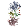
| ||||||||
|---|---|---|---|---|---|---|---|---|---|
| 1 | 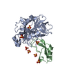
| ||||||||
| 2 | 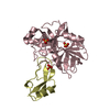
| ||||||||
| 3 | 
| ||||||||
| 4 |
| ||||||||
| 5 | 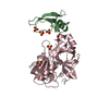
| ||||||||
| 6 | 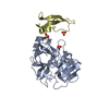
| ||||||||
| Unit cell |
|
-Components
| #1: Protein | Mass: 25686.037 Da / Num. of mol.: 2 / Mutation: K50W, M87L / Source method: isolated from a natural source / Source: (natural) #2: Protein | Mass: 6566.560 Da / Num. of mol.: 2 Source method: isolated from a genetically manipulated source Source: (gene. exp.)  #3: Chemical | ChemComp-SO4 /   Mass: 96.063 Da / Num. of mol.: 10 / Source method: obtained synthetically / Formula: SO4 Mass: 96.063 Da / Num. of mol.: 10 / Source method: obtained synthetically / Formula: SO4#4: Water | ChemComp-HOH / |  Mass: 18.015 Da / Num. of mol.: 539 / Source method: isolated from a natural source / Formula: H2O Mass: 18.015 Da / Num. of mol.: 539 / Source method: isolated from a natural source / Formula: H2OHas protein modification | Y | |
|---|
-Experimental details
-Experiment
| Experiment | Method: X-RAY DIFFRACTION / Number of used crystals: 1 |
|---|
- Sample preparation
| Crystal | Density Matthews: 4.7 Å3/Da / Density % sol: 73.4 % Description: The author notes that the R merge value noted here is a multiplicity weighted R meas |
|---|---|
| Crystal grow | Temperature: 298 K / Method: vapor diffusion, hanging drop / pH: 7.8 Details: 50% AMMONIUM SULFATE, 0.1M TRIS, pH 7.80, VAPOR DIFFUSION, HANGING DROP, temperature 298K |
-Data collection
| Diffraction | Mean temperature: 100 K |
|---|---|
| Diffraction source | Source: SYNCHROTRON / Site: ESRF  / Beamline: ID14-4 / Wavelength: 0.9312 Å / Beamline: ID14-4 / Wavelength: 0.9312 Å |
| Detector | Type: ADSC QUANTUM 4 / Detector: CCD / Date: Jun 19, 1999 |
| Radiation | Protocol: SINGLE WAVELENGTH / Monochromatic (M) / Laue (L): M / Scattering type: x-ray |
| Radiation wavelength | Wavelength: 0.9312 Å / Relative weight: 1 |
| Reflection | Resolution: 1.7→25 Å / Num. all: 125708 / Num. obs: 125561 / % possible obs: 98.8 % / Observed criterion σ(I): 0 / Redundancy: 2.7 % / Biso Wilson estimate: 22.9 Å2 / Rmerge(I) obs: 0.069 / Rsym value: 0.056 / Net I/σ(I): 7.1 |
| Reflection shell | Resolution: 1.7→1.79 Å / Redundancy: 2.3 % / Rmerge(I) obs: 0.325 / Mean I/σ(I) obs: 2.1 / Num. unique all: 17621 / Rsym value: 0.255 / % possible all: 98.8 |
- Processing
| Software |
| ||||||||||||||||||||||||||||||||||||||||||||||||||||||||||||||||||||||||||||||||
|---|---|---|---|---|---|---|---|---|---|---|---|---|---|---|---|---|---|---|---|---|---|---|---|---|---|---|---|---|---|---|---|---|---|---|---|---|---|---|---|---|---|---|---|---|---|---|---|---|---|---|---|---|---|---|---|---|---|---|---|---|---|---|---|---|---|---|---|---|---|---|---|---|---|---|---|---|---|---|---|---|---|
| Refinement | Method to determine structure: MOLECULAR REPLACEMENT Starting model: PDB ENTRY 1P2N Resolution: 1.7→14.98 Å / Rfactor Rfree error: 0.004 / Isotropic thermal model: RESTRAINED / Cross valid method: THROUGHOUT / σ(F): 0 / Stereochemistry target values: Engh & Huber
| ||||||||||||||||||||||||||||||||||||||||||||||||||||||||||||||||||||||||||||||||
| Solvent computation | Solvent model: FLAT MODEL / Bsol: 62.937 Å2 / ksol: 0.421303 e/Å3 | ||||||||||||||||||||||||||||||||||||||||||||||||||||||||||||||||||||||||||||||||
| Displacement parameters | Biso mean: 25.9 Å2
| ||||||||||||||||||||||||||||||||||||||||||||||||||||||||||||||||||||||||||||||||
| Refine analyze |
| ||||||||||||||||||||||||||||||||||||||||||||||||||||||||||||||||||||||||||||||||
| Refinement step | Cycle: LAST / Resolution: 1.7→14.98 Å
| ||||||||||||||||||||||||||||||||||||||||||||||||||||||||||||||||||||||||||||||||
| Refine LS restraints |
| ||||||||||||||||||||||||||||||||||||||||||||||||||||||||||||||||||||||||||||||||
| LS refinement shell | Resolution: 1.7→1.81 Å / Rfactor Rfree error: 0.011 / Total num. of bins used: 6
| ||||||||||||||||||||||||||||||||||||||||||||||||||||||||||||||||||||||||||||||||
| Xplor file |
|
