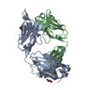
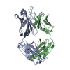
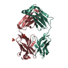
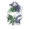
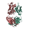
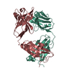
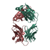
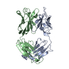
Evidence: gel filtration, fAb fragment of full length mAb
Type
Name
Symmetry operation
Number
identity operation
1_555
x,y,z
1
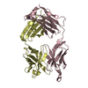
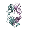
Buried area
3880 Å2
ΔGint
-77 kcal/mol
Surface area
19470 Å2
Method
PISA
Unit cell
Length a, b, c (Å)
132.018, 58.014, 80.442
Angle α, β, γ (deg.)
90.00, 124.38, 90.00
Int Tables number
5
Space group name H-M
C121
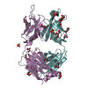
Components on special symmetry positions
ID
Model
Components
1
1
L-302-
ZN
2
1
H-301-
ZN
-
Components
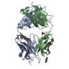
#1: Antibody
fAbLightChain
Mass: 24089.898 Da / Num. of mol.: 1 Source method: isolated from a genetically manipulated source Source: (gene. exp.) Homo sapiens (human) / Production host: Homo sapiens (human)
#2: Antibody
fAbHeavyChain
Mass: 22923.631 Da / Num. of mol.: 1 Source method: isolated from a genetically manipulated source Source: (gene. exp.) Homo sapiens (human) / Production host: Homo sapiens (human)
 Movie
Movie Controller

 Yorodumi
 Open data
 Basic information
 Components
 Keywords
 Function and homology information
 Homo sapiens (human)
 X-RAY DIFFRACTION /
  Authors
 Citation
 Structure visualization
 Downloads & links
 Other downloads
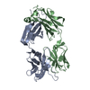


 PDBj
PDBj

 Assembly
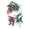

 Mass: 65.409 Da / Num. of mol.: 4 / Source method: obtained synthetically / Formula: Zn
 Mass: 92.094 Da / Num. of mol.: 1 / Source method: obtained synthetically / Formula: C3H8O3
 Mass: 18.015 Da / Num. of mol.: 47 / Source method: isolated from a natural source / Formula: H2O
 Sample preparation
 / Beamline: 21-ID-G / Wavelength: 0.97856 Å
 Processing