 Movie
Movie Controller


+ Open data

- Basic information
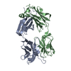
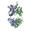
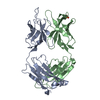
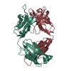
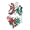
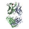
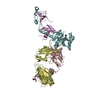
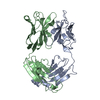
| Entry | Database: PDB / ID: 5ibu | ||||||
|---|---|---|---|---|---|---|---|
| Title | 6652 Fab (unbound) | ||||||
 Components Components |
| ||||||
 Keywords Keywords | IMMUNE SYSTEM / antibody / influenza / unbound | ||||||
| Function / homology | Immunoglobulins / Immunoglobulin-like / Sandwich / Mainly Beta Function and homology information Function and homology information | ||||||
| Biological species |  Homo sapiens (human) Homo sapiens (human) | ||||||
| Method |  X-RAY DIFFRACTION / SYNCHROTRON / MOLECULAR REPLACEMENT / molecular replacement / Resolution: 1.71 Å X-RAY DIFFRACTION / SYNCHROTRON / MOLECULAR REPLACEMENT / molecular replacement / Resolution: 1.71 Å | ||||||
 Authors Authors | Raymond, D.D. / Harrison, S.C. | ||||||
| Funding support |  United States, 1items United States, 1items
| ||||||
 Citation Citation | Journal: Nat. Med. / Year: 2016 Title: Influenza immunization elicits antibodies specific for an egg-adapted vaccine strain. Authors: Raymond, D.D. / Stewart, S.M. / Lee, J. / Ferdman, J. / Bajic, G. / Do, K.T. / Ernandes, M.J. / Suphaphiphat, P. / Settembre, E.C. / Dormitzer, P.R. / Del Giudice, G. / Finco, O. / Kang, T.H. ...Authors: Raymond, D.D. / Stewart, S.M. / Lee, J. / Ferdman, J. / Bajic, G. / Do, K.T. / Ernandes, M.J. / Suphaphiphat, P. / Settembre, E.C. / Dormitzer, P.R. / Del Giudice, G. / Finco, O. / Kang, T.H. / Ippolito, G.C. / Georgiou, G. / Kepler, T.B. / Haynes, B.F. / Moody, M.A. / Liao, H.X. / Schmidt, A.G. / Harrison, S.C. | ||||||
| History |
|
- Structure visualization
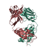
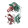
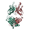
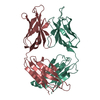
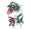
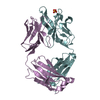
| Structure viewer | Molecule: MolmilJmol/JSmol |
|---|
- Downloads & links
-Download
| PDBx/mmCIF format | 5ibu.cif.gz | 203.8 KB | Display | PDBx/mmCIF format |
|---|---|---|---|---|
| PDB format | pdb5ibu.ent.gz | 159.3 KB | Display | PDB format |
| PDBx/mmJSON format | 5ibu.json.gz | Tree view | PDBx/mmJSON format | |
| Others |  Other downloads Other downloads |
-Validation report
| Arichive directory | https://data.pdbj.org/pub/pdb/validation_reports/ib/5ibuftp://data.pdbj.org/pub/pdb/validation_reports/ib/5ibu | HTTPS FTP |
|---|
-Related structure data
| Related structure data | 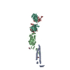 5iblC  5ibtC 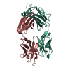 4fqlS  4hp0S C: citing same article ( S: Starting model for refinement |
|---|---|
| Similar structure data |


-Links
 PDBj
PDBj

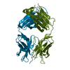
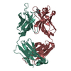
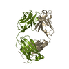
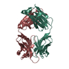
- Assembly
| Deposited unit | 
| ||||||||
|---|---|---|---|---|---|---|---|---|---|
| 1 | 
| ||||||||
| 2 | 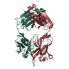
| ||||||||
| Unit cell |
|
-Components
| #1: Antibody | Mass: 23613.191 Da / Num. of mol.: 2 Source method: isolated from a genetically manipulated source Source: (gene. exp.) Homo sapiens (human) / Production host: Homo sapiens (human)#2: Antibody | Mass: 24656.707 Da / Num. of mol.: 2 Source method: isolated from a genetically manipulated source Source: (gene. exp.) Homo sapiens (human) / Production host: Homo sapiens (human)#3: Water | ChemComp-HOH / |  Mass: 18.015 Da / Num. of mol.: 1065 / Source method: isolated from a natural source / Formula: H2O Mass: 18.015 Da / Num. of mol.: 1065 / Source method: isolated from a natural source / Formula: H2OHas protein modification | Y | |
|---|
-Experimental details
-Experiment
| Experiment | Method: X-RAY DIFFRACTION / Number of used crystals: 1 |
|---|
- Sample preparation
| Crystal | Density Matthews: 2.65 Å3/Da / Density % sol: 53.6 % |
|---|---|
| Crystal grow | Temperature: 293.15 K / Method: vapor diffusion, hanging drop / pH: 5.6 Details: 0.1 M NaCitrate pH 5.6, 20% PEG 4K, 20% Isopropanol and 10% glycerol as a cryoprotectant |
-Data collection
| Diffraction | Mean temperature: 173.15 K | ||||||||||||||||||||||||||||||||||||||||||||||||||
|---|---|---|---|---|---|---|---|---|---|---|---|---|---|---|---|---|---|---|---|---|---|---|---|---|---|---|---|---|---|---|---|---|---|---|---|---|---|---|---|---|---|---|---|---|---|---|---|---|---|---|---|
| Diffraction source | Source: SYNCHROTRON / Site: ALS / Beamline: 8.2.2 / Wavelength: 0.999973 Å | ||||||||||||||||||||||||||||||||||||||||||||||||||
| Detector | Type: ADSC QUANTUM 315 / Detector: CCD / Date: Sep 26, 2013 | ||||||||||||||||||||||||||||||||||||||||||||||||||
| Radiation | Protocol: SINGLE WAVELENGTH / Monochromatic (M) / Laue (L): M / Scattering type: x-ray | ||||||||||||||||||||||||||||||||||||||||||||||||||
| Radiation wavelength | Wavelength: 0.999973 Å / Relative weight: 1 | ||||||||||||||||||||||||||||||||||||||||||||||||||
| Reflection | Resolution: 1.71→44.78 Å / Num. obs: 103494 / % possible obs: 95.1 % / Observed criterion σ(I): -3 / Redundancy: 3.1 % / Biso Wilson estimate: 25.65 Å2 / CC1/2: 0.994 / Rmerge(I) obs: 0.112 / Net I/σ(I): 11.88 | ||||||||||||||||||||||||||||||||||||||||||||||||||
| Reflection shell |
|
-Phasing
| Phasing | Method: molecular replacement |
|---|
- Processing
| Software |
| ||||||||||||||||||||||||||||||||||||||||||||||||||||||||||||||||||||||||||||||||||||||||||||||||||||||||||||
|---|---|---|---|---|---|---|---|---|---|---|---|---|---|---|---|---|---|---|---|---|---|---|---|---|---|---|---|---|---|---|---|---|---|---|---|---|---|---|---|---|---|---|---|---|---|---|---|---|---|---|---|---|---|---|---|---|---|---|---|---|---|---|---|---|---|---|---|---|---|---|---|---|---|---|---|---|---|---|---|---|---|---|---|---|---|---|---|---|---|---|---|---|---|---|---|---|---|---|---|---|---|---|---|---|---|---|---|---|---|
| Refinement | Method to determine structure: MOLECULAR REPLACEMENT Starting model: 4HP0,4FQL Resolution: 1.71→44.78 Å / Cor.coef. Fo:Fc: 0.939 / Cor.coef. Fo:Fc free: 0.92 / Rfactor Rfree error: 0 / SU R Cruickshank DPI: 0.119 / Cross valid method: THROUGHOUT / σ(F): 0 / SU R Blow DPI: 0.128 / SU Rfree Blow DPI: 0.121 / SU Rfree Cruickshank DPI: 0.116
| ||||||||||||||||||||||||||||||||||||||||||||||||||||||||||||||||||||||||||||||||||||||||||||||||||||||||||||
| Displacement parameters | Biso max: 116.43 Å2 / Biso mean: 25.9 Å2 / Biso min: 10.18 Å2
| ||||||||||||||||||||||||||||||||||||||||||||||||||||||||||||||||||||||||||||||||||||||||||||||||||||||||||||
| Refine analyze | Luzzati coordinate error obs: 0.27 Å | ||||||||||||||||||||||||||||||||||||||||||||||||||||||||||||||||||||||||||||||||||||||||||||||||||||||||||||
| Refinement step | Cycle: final / Resolution: 1.71→44.78 Å
| ||||||||||||||||||||||||||||||||||||||||||||||||||||||||||||||||||||||||||||||||||||||||||||||||||||||||||||
| Refine LS restraints |
| ||||||||||||||||||||||||||||||||||||||||||||||||||||||||||||||||||||||||||||||||||||||||||||||||||||||||||||
| LS refinement shell | Resolution: 1.71→1.75 Å / Total num. of bins used: 20
|
