 Movie
Movie Controller

[English] 日本語
 Yorodumi
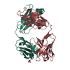
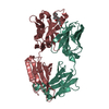
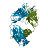
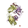
- PDB-1ggi: CRYSTAL STRUCTURE OF AN HIV-1 NEUTRALIZING ANTIBODY 50.1 IN COMPL... -
+ Open data

- Basic information
| Entry | Database: PDB / ID: 1ggi | ||||||
|---|---|---|---|---|---|---|---|
| Title | CRYSTAL STRUCTURE OF AN HIV-1 NEUTRALIZING ANTIBODY 50.1 IN COMPLEX WITH ITS V3 LOOP PEPTIDE ANTIGEN | ||||||
 Components Components |
| ||||||
 Keywords Keywords | IMMUNOGLOBULIN | ||||||
| Function / homology |  Function and homology information Function and homology informationFc-gamma receptor I complex binding / immunoglobulin complex, circulating / immunoglobulin receptor binding / IgG immunoglobulin complex / Dectin-2 family / complement activation, classical pathway / antigen binding / symbiont-mediated perturbation of host defense response / positive regulation of plasma membrane raft polarization / positive regulation of receptor clustering ...Fc-gamma receptor I complex binding / immunoglobulin complex, circulating / immunoglobulin receptor binding / IgG immunoglobulin complex / Dectin-2 family / complement activation, classical pathway / antigen binding / symbiont-mediated perturbation of host defense response / positive regulation of plasma membrane raft polarization / positive regulation of receptor clustering / host cell endosome membrane / antibacterial humoral response / clathrin-dependent endocytosis of virus by host cell / viral protein processing / fusion of virus membrane with host plasma membrane / fusion of virus membrane with host endosome membrane / viral envelope / virion attachment to host cell / host cell plasma membrane / virion membrane / structural molecule activity / : / membrane / identical protein binding Similarity search - Function | ||||||
| Biological species |  | ||||||
| Method |  X-RAY DIFFRACTION / Resolution: 2.8 Å X-RAY DIFFRACTION / Resolution: 2.8 Å | ||||||
 Authors Authors | Stanfield, R.L. / Rini, J.M. / Wilson, I.A. | ||||||
 Citation Citation | Journal: Proc.Natl.Acad.Sci.USA / Year: 1993 Title: Crystal structure of a human immunodeficiency virus type 1 neutralizing antibody, 50.1, in complex with its V3 loop peptide antigen. Authors: Rini, J.M. / Stanfield, R.L. / Stura, E.A. / Salinas, P.A. / Profy, A.T. / Wilson, I.A. #1: Journal: Proteins / Year: 1992Title: Crystallization, Sequence, and Preliminary Crystallographic Data for an Antipeptide Fab 50.1 And Peptide Complexes with the Principal Neutralizing Determinant of HIV-1 Gp120 Authors: Stura, E.A. / Stanfield, R.L. / Fieser, G.G. / Silver, S. / Rogusca, M. / Hincapie, L.M. / Simmerman, H.K.B. / Profy, A.T. / Wilson, I.A. #2: Journal: To be PublishedTitle: Major Antigen-Induced Domain Rearrangements in an Antibody Authors: Stanfield, R.L. / Takimoto-Kamimura, M. / Rini, J.M. / Profy, A.T. / Wilson, I.A. | ||||||
| History |
|
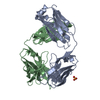
- Structure visualization
| Structure viewer | Molecule: MolmilJmol/JSmol |
|---|
- Downloads & links
-Download
| PDBx/mmCIF format | 1ggi.cif.gz | 162.9 KB | Display | PDBx/mmCIF format |
|---|---|---|---|---|
| PDB format | pdb1ggi.ent.gz | 128.6 KB | Display | PDB format |
| PDBx/mmJSON format | 1ggi.json.gz | Tree view | PDBx/mmJSON format | |
| Others |  Other downloads Other downloads |
-Validation report
| Arichive directory | https://data.pdbj.org/pub/pdb/validation_reports/gg/1ggiftp://data.pdbj.org/pub/pdb/validation_reports/gg/1ggi | HTTPS FTP |
|---|
-Related structure data
| Similar structure data |
|---|


-Links
 PDBj
PDBj


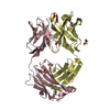
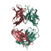
- Assembly
| Deposited unit | 
| ||||||||||||||||||||
|---|---|---|---|---|---|---|---|---|---|---|---|---|---|---|---|---|---|---|---|---|---|
| 1 | 
| ||||||||||||||||||||
| 2 | 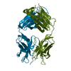
| ||||||||||||||||||||
| Unit cell |
| ||||||||||||||||||||
| Atom site foot note | 1: RESIDUES 8, 77, 95, 141, 204 OF THE L AND M CHAINS, AND RESIDUES 149, 151, 200 OF THE H AND J CHAINS ARE CIS PROLINES. | ||||||||||||||||||||
| Noncrystallographic symmetry (NCS) | NCS oper:
| ||||||||||||||||||||
| Details | THERE ARE TWO FAB MOLECULES IN THE ASYMMETRIC UNIT. THE TRANSFORMATIONS GIVEN ON *MTRIX* RECORDS BELOW YIELD APPROXIMATE COORDINATES FOR CHAINS *L* AND *H* WHEN APPLIED TO CHAINS *M* AND *J*. THESE TRANSFORMATIONS HAVE BEEN PROVIDED SEPARATELY FOR THE VH AND CH DOMAINS OF THE HEAVY CHAIN AND THE VL AND CL DOMAINS OF THE LIGHT CHAIN. THE TRANSFORMATION PRESENTED ON *MTRIX 1* RECORDS BELOW WILL YIELD APPROXIMATE COORDINATES FOR THE VL DOMAIN OF CHAIN *L* WHEN APPLIED TO THE VL DOMAIN OF CHAIN *M*. THE TRANSFORMATION PRESENTED ON *MTRIX 2* RECORDS BELOW WILL YIELD APPROXIMATE COORDINATES FOR THE CL DOMAIN OF CHAIN *L* WHEN APPLIED TO THE CL DOMAIN OF CHAIN *M*. THE TRANSFORMATION PRESENTED ON *MTRIX 3* RECORDS BELOW WILL YIELD APPROXIMATE COORDINATES FOR THE VH DOMAIN OF CHAIN *H* WHEN APPLIED TO THE VH DOMAIN OF CHAIN *J*. THE TRANSFORMATION PRESENTED ON *MTRIX 4* RECORDS BELOW WILL YIELD APPROXIMATE COORDINATES FOR THE CH DOMAIN OF CHAIN *H* WHEN APPLIED TO THE CH DOMAIN OF CHAIN *J*. |
-Components
| #1: Antibody | Mass: 23994.336 Da / Num. of mol.: 2 Source method: isolated from a genetically manipulated source Source: (gene. exp.) #2: Antibody | Mass: 23828.807 Da / Num. of mol.: 2 Source method: isolated from a genetically manipulated source References: UniProt: P01863 #3: Protein/peptide | Mass: 1827.182 Da / Num. of mol.: 2 Source method: isolated from a genetically manipulated source References: UniProt: P05877*PLUS Has protein modification | Y | Sequence details | THE FAB FRAGMENT IS NUMBERED BY THE CONVENTION OF E. KABAT (E.A. KABAT, T.T. WU, M. REID-MILLER, H. ...THE FAB FRAGMENT IS NUMBERED BY THE CONVENTION | |
|---|
-Experimental details
-Experiment
| Experiment | Method: X-RAY DIFFRACTION |
|---|
- Sample preparation
| Crystal | Density Matthews: 2.8 Å3/Da / Density % sol: 56.14 % | |||||||||||||||||||||||||
|---|---|---|---|---|---|---|---|---|---|---|---|---|---|---|---|---|---|---|---|---|---|---|---|---|---|---|
| Crystal grow | *PLUS pH: 5.5 / Method: vapor diffusion | |||||||||||||||||||||||||
| Components of the solutions | *PLUS
|
-Data collection
| Radiation | Scattering type: x-ray |
|---|---|
| Radiation wavelength | Relative weight: 1 |
| Reflection | *PLUS Highest resolution: 2.8 Å / % possible obs: 81 % / Rmerge(I) obs: 0.1 |
- Processing
| Software |
| ||||||||||||||||||||||||||||||||||||||||||||||||||||||||||||
|---|---|---|---|---|---|---|---|---|---|---|---|---|---|---|---|---|---|---|---|---|---|---|---|---|---|---|---|---|---|---|---|---|---|---|---|---|---|---|---|---|---|---|---|---|---|---|---|---|---|---|---|---|---|---|---|---|---|---|---|---|---|
| Refinement | Rfactor Rwork: 0.188 / Rfactor obs: 0.188 / Highest resolution: 2.8 Å | ||||||||||||||||||||||||||||||||||||||||||||||||||||||||||||
| Refinement step | Cycle: LAST / Highest resolution: 2.8 Å
| ||||||||||||||||||||||||||||||||||||||||||||||||||||||||||||
| Refine LS restraints |
| ||||||||||||||||||||||||||||||||||||||||||||||||||||||||||||
| Refinement | *PLUS Highest resolution: 2.8 Å / Lowest resolution: 10 Å / Rfactor obs: 0.188 | ||||||||||||||||||||||||||||||||||||||||||||||||||||||||||||
| Solvent computation | *PLUS | ||||||||||||||||||||||||||||||||||||||||||||||||||||||||||||
| Displacement parameters | *PLUS | ||||||||||||||||||||||||||||||||||||||||||||||||||||||||||||
| Refine LS restraints | *PLUS Type: x_angle_d / Dev ideal: 3.79 |
