 Movie
Movie Controller

[English] 日本語
 Yorodumi
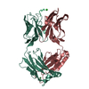
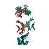
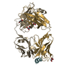
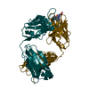
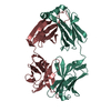
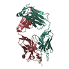
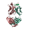
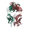
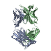
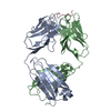
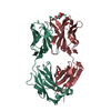
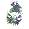
- PDB-1e4x: crossreactive binding of a circularized peptide to an anti-TGFalp... -
+ Open data

- Basic information
| Entry | Database: PDB / ID: 1e4x | ||||||
|---|---|---|---|---|---|---|---|
| Title | crossreactive binding of a circularized peptide to an anti-TGFalpha antibody Fab-fragment | ||||||
 Components Components |
| ||||||
 Keywords Keywords | IMMUNE SYSTEM / COMPLEX (ANTIBODY-ANTIGEN) / CROSS-REACTIVITY / PROTEIN-PEPTIDE RECOGNITION | ||||||
| Function / homology |  Function and homology information Function and homology informationalpha-beta T cell receptor complex / IgG immunoglobulin complex / B cell differentiation / adaptive immune response / extracellular region / plasma membrane Similarity search - Function | ||||||
| Biological species |  SYNTHETIC CONSTRUCT (others) | ||||||
| Method |  X-RAY DIFFRACTION / SYNCHROTRON / MOLECULAR REPLACEMENT / Resolution: 1.9 Å X-RAY DIFFRACTION / SYNCHROTRON / MOLECULAR REPLACEMENT / Resolution: 1.9 Å | ||||||
 Authors Authors | Hahn, M. / Winkler, D. / Misselwitz, R. / Wessner, H. / Welfle, K. / Zahn, G. / Schneider-Mergener, J. / Hoehne, W. | ||||||
 Citation Citation | Journal: J.Mol.Biol. / Year: 2001 Title: Cross-Reactive Binding of Cyclic Peptides to an Anti-Tgf Alpha Antibody Fab Fragment: An X-Ray Structural and Thermodynamic Analysis Authors: Hahn, M. / Winkler, D. / Welfle, K. / Misselwitz, R. / Welfle, H. / Wessner, H. / Zahn, G. / Scholz, C. / Seifert, M. / Harkins, R. / Schneider-Mergener, J. / Hoehne, W. | ||||||
| History |
|
- Structure visualization
| Structure viewer | Molecule: MolmilJmol/JSmol |
|---|
- Downloads & links
-Download
| PDBx/mmCIF format | 1e4x.cif.gz | 185 KB | Display | PDBx/mmCIF format |
|---|---|---|---|---|
| PDB format | pdb1e4x.ent.gz | 146.6 KB | Display | PDB format |
| PDBx/mmJSON format | 1e4x.json.gz | Tree view | PDBx/mmJSON format | |
| Others |  Other downloads Other downloads |
-Validation report
| Arichive directory | https://data.pdbj.org/pub/pdb/validation_reports/e4/1e4xftp://data.pdbj.org/pub/pdb/validation_reports/e4/1e4x | HTTPS FTP |
|---|
-Related structure data


-Links
 PDBj
PDBj


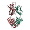
- Assembly
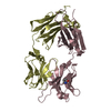
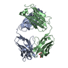
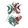
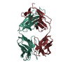
| Deposited unit | 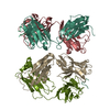
| ||||||||
|---|---|---|---|---|---|---|---|---|---|
| 1 | 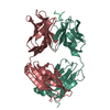
| ||||||||
| 2 | 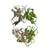
| ||||||||
| Unit cell |
| ||||||||
| Noncrystallographic symmetry (NCS) | NCS oper: (Code: given Matrix: (0.23405, -0.18859, -0.95376), Vector: |
-Components
| #1: Antibody | Mass: 23351.205 Da / Num. of mol.: 1 / Fragment: IG KAPPA HEAVY CHAIN / Source method: isolated from a natural source / Details: MURINE FAB-FRAGMENT / Source: (natural) | ||||||||
|---|---|---|---|---|---|---|---|---|---|
| #2: Antibody | Mass: 23377.242 Da / Num. of mol.: 1 / Fragment: IG KAPPA HEAVY CHAIN / Source method: isolated from a natural source / Details: MURINE FAB-FRAGMENT / Source: (natural) | ||||||||
| #3: Antibody | Mass: 23577.947 Da / Num. of mol.: 2 / Fragment: IG KAPPA LIGHT CHAIN / Source method: isolated from a natural source / Details: MURINE FAB-FRAGMENT / Source: (natural) #4: Protein/peptide | Mass: 817.866 Da / Num. of mol.: 2 / Source method: obtained synthetically / Source: (synth.) SYNTHETIC CONSTRUCT (others) #5: Water | ChemComp-HOH / |  Mass: 18.015 Da / Num. of mol.: 407 / Source method: isolated from a natural source / Formula: H2O Mass: 18.015 Da / Num. of mol.: 407 / Source method: isolated from a natural source / Formula: H2OCompound details | CHAIN P IS A EPITOPE PEPTIDE WITH THE SEQUENCE VAL-VAL-SER-HIS-PHE-ASN-ASP, DERIVED FROM THE N- ...CHAIN P IS A EPITOPE PEPTIDE WITH THE SEQUENCE VAL-VAL-SER-HIS-PHE-ASN-ASP, DERIVED FROM THE N-TERMINUS OF TGFALPHA. CHAIN L, M: THE C-TERMINAL REGION IS THE CONSTANT REGION OF THE IG KAPPA LIGHT CHAIN. THE N-TERMINAL REGION IS THE VARIABLE REGION OF THE IG KAPPA LIGHT CHAIN. THE VARIABLE REGION IS AUTOANTIBO | Has protein modification | Y | |
-Experimental details
-Experiment
| Experiment | Method: X-RAY DIFFRACTION / Number of used crystals: 1 |
|---|
- Sample preparation
| Crystal | Density Matthews: 2.5 Å3/Da / Density % sol: 40.7 % | ||||||||||||||||||||
|---|---|---|---|---|---|---|---|---|---|---|---|---|---|---|---|---|---|---|---|---|---|
| Crystal grow | pH: 8.5 Details: PROTEIN IN TRIS-HCL, 8.5, 6-10 MG/ML PRECIPITANT: 12% PEG 8000, 20 MM NA-ACETATE, pH 8.50 | ||||||||||||||||||||
| Crystal grow | *PLUS Method: vapor diffusion, hanging drop | ||||||||||||||||||||
| Components of the solutions | *PLUS
|
-Data collection
| Diffraction | Mean temperature: 100 K |
|---|---|
| Diffraction source | Source: SYNCHROTRON / Site: MPG/DESY, HAMBURG  / Beamline: BW6 / Wavelength: 1.3 / Beamline: BW6 / Wavelength: 1.3 |
| Detector | Type: XRAY RESEARCH / Detector: CCD / Date: Jun 17, 1999 |
| Radiation | Protocol: SINGLE WAVELENGTH / Monochromatic (M) / Laue (L): M / Scattering type: x-ray |
| Radiation wavelength | Wavelength: 1.3 Å / Relative weight: 1 |
| Reflection | Resolution: 1.9→20 Å / Num. obs: 68086 / % possible obs: 92.9 % / Redundancy: 5.2 % / Biso Wilson estimate: 22.7 Å2 / Rmerge(I) obs: 0.038 / Net I/σ(I): 29.7 |
| Reflection shell | Resolution: 1.9→1.97 Å / Redundancy: 1.7 % / Rmerge(I) obs: 0.257 / Mean I/σ(I) obs: 2.4 / % possible all: 82.8 |
| Reflection | *PLUS Lowest resolution: 20 Å |
| Reflection shell | *PLUS % possible obs: 82.8 % / Mean I/σ(I) obs: 2.2 |
- Processing
| Software |
| ||||||||||||||||||||||||||||||||||||||||||||||||||||||||||||||||||||||||||||||||||||
|---|---|---|---|---|---|---|---|---|---|---|---|---|---|---|---|---|---|---|---|---|---|---|---|---|---|---|---|---|---|---|---|---|---|---|---|---|---|---|---|---|---|---|---|---|---|---|---|---|---|---|---|---|---|---|---|---|---|---|---|---|---|---|---|---|---|---|---|---|---|---|---|---|---|---|---|---|---|---|---|---|---|---|---|---|---|
| Refinement | Method to determine structure: MOLECULAR REPLACEMENT Starting model: TAB2 Resolution: 1.9→20 Å / Cross valid method: THROUGHOUT / σ(F): 0
| ||||||||||||||||||||||||||||||||||||||||||||||||||||||||||||||||||||||||||||||||||||
| Displacement parameters | Biso mean: 36 Å2 | ||||||||||||||||||||||||||||||||||||||||||||||||||||||||||||||||||||||||||||||||||||
| Refinement step | Cycle: LAST / Resolution: 1.9→20 Å
| ||||||||||||||||||||||||||||||||||||||||||||||||||||||||||||||||||||||||||||||||||||
| Refine LS restraints |
| ||||||||||||||||||||||||||||||||||||||||||||||||||||||||||||||||||||||||||||||||||||
| Software | *PLUS Name: REFMAC / Classification: refinement | ||||||||||||||||||||||||||||||||||||||||||||||||||||||||||||||||||||||||||||||||||||
| Refinement | *PLUS Lowest resolution: 20 Å / Rfactor obs: 0.245 | ||||||||||||||||||||||||||||||||||||||||||||||||||||||||||||||||||||||||||||||||||||
| Solvent computation | *PLUS | ||||||||||||||||||||||||||||||||||||||||||||||||||||||||||||||||||||||||||||||||||||
| Displacement parameters | *PLUS |
