 Movie
Movie Controller

[English] 日本語
 Yorodumi
- PDB-4xh2: Crystal structure of human paxillin LD4 motif in complex with Fab... -
+ Open data

- Basic information
| Entry | Database: PDB / ID: 4xh2 | ||||||
|---|---|---|---|---|---|---|---|
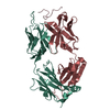
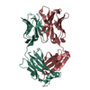
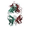
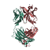
| Title | Crystal structure of human paxillin LD4 motif in complex with Fab fragment | ||||||
 Components Components |
| ||||||
 Keywords Keywords | CELL ADHESION / synthetic antibody / paxillin / LD motif / immunoglobulin / Fab fragment / complex / focal adhesion | ||||||
| Function / homology |  Function and homology information Function and homology informationRegulation of cytoskeletal remodeling and cell spreading by IPP complex components / Localization of the PINCH-ILK-PARVIN complex to focal adhesions / Regulation of MITF-M-dependent genes involved in extracellular matrix, focal adhesion and epithelial-to-mesenchymal transition / neuropilin binding / vinculin binding / signal complex assembly / microtubule associated complex / growth hormone receptor signaling pathway / Smooth Muscle Contraction / GAB1 signalosome ...Regulation of cytoskeletal remodeling and cell spreading by IPP complex components / Localization of the PINCH-ILK-PARVIN complex to focal adhesions / Regulation of MITF-M-dependent genes involved in extracellular matrix, focal adhesion and epithelial-to-mesenchymal transition / neuropilin binding / vinculin binding / signal complex assembly / microtubule associated complex / growth hormone receptor signaling pathway / Smooth Muscle Contraction / GAB1 signalosome / endothelial cell migration / positive regulation of stress fiber assembly / substrate adhesion-dependent cell spreading / PTK6 Regulates RHO GTPases, RAS GTPase and MAP kinases / stress fiber / transforming growth factor beta receptor signaling pathway / cellular response to reactive oxygen species / beta-catenin binding / VEGFA-VEGFR2 Pathway / cell-cell junction / cell migration / lamellipodium / protein phosphatase binding / cell cortex / cell adhesion / focal adhesion / signal transduction / metal ion binding / plasma membrane / cytosol Similarity search - Function | ||||||
| Biological species |  Homo sapiens (human) Homo sapiens (human)synthetic construct (others) | ||||||
| Method |  X-RAY DIFFRACTION / SYNCHROTRON / MOLECULAR REPLACEMENT / Resolution: 2 Å X-RAY DIFFRACTION / SYNCHROTRON / MOLECULAR REPLACEMENT / Resolution: 2 Å | ||||||
 Authors Authors | Nocula-Lugowska, M. / Lugowski, M. / Salgia, R. / Kossiakoff, A.A. | ||||||
| Funding support |  United States, 1items United States, 1items
| ||||||
 Citation Citation | Journal: J.Mol.Biol. / Year: 2015 Title: Engineering Synthetic Antibody Inhibitors Specific for LD2 or LD4 Motifs of Paxillin. Authors: Nocula-Lugowska, M. / Lugowski, M. / Salgia, R. / Kossiakoff, A.A. | ||||||
| History |
|
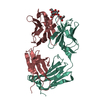
- Structure visualization
| Structure viewer | Molecule: MolmilJmol/JSmol |
|---|
- Downloads & links
-Download
| PDBx/mmCIF format | 4xh2.cif.gz | 535.2 KB | Display | PDBx/mmCIF format |
|---|---|---|---|---|
| PDB format | pdb4xh2.ent.gz | 439.8 KB | Display | PDB format |
| PDBx/mmJSON format | 4xh2.json.gz | Tree view | PDBx/mmJSON format | |
| Others |  Other downloads Other downloads |
-Validation report
| Arichive directory | https://data.pdbj.org/pub/pdb/validation_reports/xh/4xh2ftp://data.pdbj.org/pub/pdb/validation_reports/xh/4xh2 | HTTPS FTP |
|---|
-Related structure data
| Related structure data | 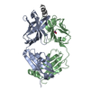 4xgzC 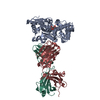 3pgfS C: citing same article ( S: Starting model for refinement |
|---|---|
| Similar structure data |
-Links
 PDBj
PDBj


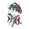
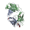
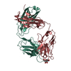
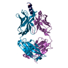
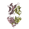
- Assembly
| Deposited unit | 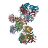
| ||||||||
|---|---|---|---|---|---|---|---|---|---|
| 1 | 
| ||||||||
| 2 | 
| ||||||||
| 3 | 
| ||||||||
| 4 | 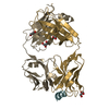
| ||||||||
| 5 | 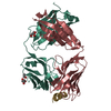
| ||||||||
| 6 | 
| ||||||||
| Unit cell |
|
-Components
-Protein/peptide , 1 types, 6 molecules aceghj
| #3: Protein/peptide | Mass: 1939.088 Da / Num. of mol.: 6 / Source method: obtained synthetically / Source: (synth.) synthetic construct (others) / References: UniProt: P49023*PLUS |
|---|
-Antibody , 2 types, 12 molecules ACEGHJBDFIKL
| #1: Antibody | Mass: 24261.975 Da / Num. of mol.: 6 Source method: isolated from a genetically manipulated source Source: (gene. exp.) Homo sapiens (human) / Details (production host): phoA promoter / Production host:  #2: Antibody | Mass: 23714.400 Da / Num. of mol.: 6 Source method: isolated from a genetically manipulated source Source: (gene. exp.) Homo sapiens (human) / Details (production host): phoA promoter / Production host: |
|---|
-Non-polymers , 6 types, 1582 molecules 


| #4: Chemical | ChemComp-GOL /  Mass: 92.094 Da / Num. of mol.: 20 / Source method: obtained synthetically / Formula: C3H8O3 Mass: 92.094 Da / Num. of mol.: 20 / Source method: obtained synthetically / Formula: C3H8O3#5: Chemical |  Mass: 94.971 Da / Num. of mol.: 2 / Source method: obtained synthetically / Formula: PO4 Mass: 94.971 Da / Num. of mol.: 2 / Source method: obtained synthetically / Formula: PO4#6: Chemical | ChemComp-ACT / |  Mass: 59.044 Da / Num. of mol.: 1 / Source method: obtained synthetically / Formula: C2H3O2 Mass: 59.044 Da / Num. of mol.: 1 / Source method: obtained synthetically / Formula: C2H3O2#7: Chemical | ChemComp-LDA / |  Mass: 229.402 Da / Num. of mol.: 1 / Source method: obtained synthetically / Formula: C14H31NO / Comment: LDAO, detergent*YM Mass: 229.402 Da / Num. of mol.: 1 / Source method: obtained synthetically / Formula: C14H31NO / Comment: LDAO, detergent*YM#8: Chemical |  Mass: 44.053 Da / Num. of mol.: 2 / Source method: obtained synthetically / Formula: C2H4O Mass: 44.053 Da / Num. of mol.: 2 / Source method: obtained synthetically / Formula: C2H4O#9: Water | ChemComp-HOH / | Mass: 18.015 Da / Num. of mol.: 1556 / Source method: isolated from a natural source / Formula: H2O |
|---|
-Experimental details
-Experiment
| Experiment | Method: X-RAY DIFFRACTION / Number of used crystals: 2 |
|---|
- Sample preparation
| Crystal | Density Matthews: 3.03 Å3/Da / Density % sol: 59.4 % |
|---|---|
| Crystal grow | Temperature: 293 K / Method: vapor diffusion, hanging drop / pH: 6.5 Details: 18% PEG 8000, 0.1 M MES, 0.5 % n-Dodecyl-N,N-Dimethylamine-N-Oxide (LDAO) |
-Data collection
| Diffraction | Mean temperature: 100 K |
|---|---|
| Diffraction source | Source: SYNCHROTRON / Site: APS / Beamline: 24-ID-C / Wavelength: 0.9792 Å |
| Detector | Type: PSI PILATUS 6M / Detector: PIXEL / Date: Oct 14, 2012 |
| Radiation | Protocol: SINGLE WAVELENGTH / Monochromatic (M) / Laue (L): M / Scattering type: x-ray |
| Radiation wavelength | Wavelength: 0.9792 Å / Relative weight: 1 |
| Reflection | Resolution: 2→117.5 Å / Num. obs: 228060 / % possible obs: 93.2 % / Observed criterion σ(I): -3 / Redundancy: 4.9 % / Biso Wilson estimate: 37.88 Å2 / Rmerge(I) obs: 0.155 / Net I/σ(I): 7.6 |
| Reflection shell | Resolution: 2→2.2 Å / Redundancy: 3.8 % / Rmerge(I) obs: 0.997 / Mean I/σ(I) obs: 1.76 / % possible all: 84.2 |
- Processing
| Software |
| ||||||||||||||||||||||||||||||||||||||||||||||||||||||||||||||||||||||||||||||||||||||||||||||||||||||||||||||||||||||||||||||||||||||||||||||||||||||||||||||||||||||||||||||||||||||
|---|---|---|---|---|---|---|---|---|---|---|---|---|---|---|---|---|---|---|---|---|---|---|---|---|---|---|---|---|---|---|---|---|---|---|---|---|---|---|---|---|---|---|---|---|---|---|---|---|---|---|---|---|---|---|---|---|---|---|---|---|---|---|---|---|---|---|---|---|---|---|---|---|---|---|---|---|---|---|---|---|---|---|---|---|---|---|---|---|---|---|---|---|---|---|---|---|---|---|---|---|---|---|---|---|---|---|---|---|---|---|---|---|---|---|---|---|---|---|---|---|---|---|---|---|---|---|---|---|---|---|---|---|---|---|---|---|---|---|---|---|---|---|---|---|---|---|---|---|---|---|---|---|---|---|---|---|---|---|---|---|---|---|---|---|---|---|---|---|---|---|---|---|---|---|---|---|---|---|---|---|---|---|---|
| Refinement | Method to determine structure: MOLECULAR REPLACEMENT Starting model: 3PGF Resolution: 2→117.5 Å / Cor.coef. Fo:Fc: 0.938 / Cor.coef. Fo:Fc free: 0.917 / SU B: 4.764 / SU ML: 0.127 / Cross valid method: THROUGHOUT / σ(F): 0 / ESU R: 0.191 / ESU R Free: 0.17 / Stereochemistry target values: MAXIMUM LIKELIHOOD / Details: HYDROGENS HAVE BEEN ADDED IN THE RIDING
| ||||||||||||||||||||||||||||||||||||||||||||||||||||||||||||||||||||||||||||||||||||||||||||||||||||||||||||||||||||||||||||||||||||||||||||||||||||||||||||||||||||||||||||||||||||||
| Solvent computation | Ion probe radii: 0.8 Å / Shrinkage radii: 0.8 Å / VDW probe radii: 1.2 Å / Solvent model: MASK | ||||||||||||||||||||||||||||||||||||||||||||||||||||||||||||||||||||||||||||||||||||||||||||||||||||||||||||||||||||||||||||||||||||||||||||||||||||||||||||||||||||||||||||||||||||||
| Displacement parameters | Biso mean: 27.33 Å2
| ||||||||||||||||||||||||||||||||||||||||||||||||||||||||||||||||||||||||||||||||||||||||||||||||||||||||||||||||||||||||||||||||||||||||||||||||||||||||||||||||||||||||||||||||||||||
| Refinement step | Cycle: LAST / Resolution: 2→117.5 Å
| ||||||||||||||||||||||||||||||||||||||||||||||||||||||||||||||||||||||||||||||||||||||||||||||||||||||||||||||||||||||||||||||||||||||||||||||||||||||||||||||||||||||||||||||||||||||
| Refine LS restraints |
|