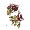
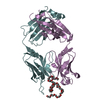
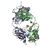
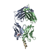
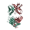
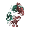
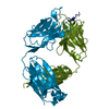
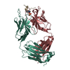
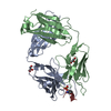
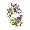
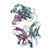
[English] 日本語
Yorodumi
- PDB-6h06: FAB CBTAU-22.1 IN COMPLEX WITH TAU PEPTIDE V1088-5 -

+
Open data


ID or keywords:

Loading...

-
Basic information

Entry
Database: PDB / ID: 6h06
TitleFAB CBTAU-22.1 IN COMPLEX WITH TAU PEPTIDE V1088-5
Components
  • HUMAN FAB ANTIBODY FRAGMENT OF CBTAU-22.1
  • HUMAN FAB ANTIBODY FRAGMENT OF HCBTAU-22.1
  • Microtubule-associated protein tauTau protein
KeywordsIMMUNE SYSTEM / FAB / Tau peptide / complex
Function / homology
Function and homology information


plus-end-directed organelle transport along microtubule / axonal transport / histone-dependent DNA binding / neurofibrillary tangle assembly / positive regulation of diacylglycerol kinase activity / negative regulation of establishment of protein localization to mitochondrion / neurofibrillary tangle / positive regulation of protein localization to synapse / microtubule lateral binding / tubulin complex ...plus-end-directed organelle transport along microtubule / axonal transport / histone-dependent DNA binding / neurofibrillary tangle assembly / positive regulation of diacylglycerol kinase activity / negative regulation of establishment of protein localization to mitochondrion / neurofibrillary tangle / positive regulation of protein localization to synapse / microtubule lateral binding / tubulin complex / phosphatidylinositol bisphosphate binding / main axon / regulation of long-term synaptic depression / negative regulation of kinase activity / negative regulation of tubulin deacetylation / generation of neurons / regulation of chromosome organization / positive regulation of protein localization / rRNA metabolic process / internal protein amino acid acetylation / regulation of mitochondrial fission / lipoprotein particle binding / intracellular distribution of mitochondria / axonal transport of mitochondrion / axon development / central nervous system neuron development / regulation of microtubule polymerization / microtubule polymerization / minor groove of adenine-thymine-rich DNA binding / negative regulation of mitochondrial membrane potential / dynactin binding / glial cell projection / apolipoprotein binding / protein polymerization / negative regulation of mitochondrial fission / axolemma / Caspase-mediated cleavage of cytoskeletal proteins / regulation of microtubule polymerization or depolymerization / positive regulation of axon extension / supramolecular fiber organization / Activation of AMPK downstream of NMDARs / cytoplasmic microtubule organization / regulation of microtubule cytoskeleton organization / stress granule assembly / regulation of cellular response to heat / regulation of calcium-mediated signaling / axon cytoplasm / positive regulation of microtubule polymerization / cellular response to brain-derived neurotrophic factor stimulus / somatodendritic compartment / synapse assembly / phosphatidylinositol binding / nuclear periphery / cellular response to nerve growth factor stimulus / positive regulation of superoxide anion generation / protein phosphatase 2A binding / regulation of autophagy / astrocyte activation / synapse organization / microglial cell activation / response to lead ion / Hsp90 protein binding / regulation of synaptic plasticity / PKR-mediated signaling / protein homooligomerization / cytoplasmic ribonucleoprotein granule / memory / microtubule cytoskeleton organization / SH3 domain binding / cellular response to reactive oxygen species / neuron projection development / activation of cysteine-type endopeptidase activity involved in apoptotic process / microtubule cytoskeleton / protein-macromolecule adaptor activity / single-stranded DNA binding / cell-cell signaling / cellular response to heat / cell body / actin binding / growth cone / protein-folding chaperone binding / double-stranded DNA binding / microtubule binding / microtubule / amyloid fibril formation / sequence-specific DNA binding / dendritic spine / learning or memory / neuron projection / nuclear speck / membrane raft / axon / negative regulation of gene expression / dendrite / neuronal cell body / DNA damage response / protein kinase binding / enzyme binding / mitochondrion / DNA binding
Similarity search - Function
: / Microtubule associated protein, tubulin-binding repeat / Microtubule-associated protein Tau / Tau and MAP protein, tubulin-binding repeat / Tau and MAP proteins tubulin-binding repeat signature. / Tau and MAP proteins tubulin-binding repeat profile. / Immunoglobulins / Immunoglobulin-like / Sandwich / Mainly Beta
Similarity search - Domain/homology
Microtubule-associated protein tau
Similarity search - Component
Biological speciesHomo sapiens (human)
MethodX-RAY DIFFRACTION / SYNCHROTRON / MOLECULAR REPLACEMENT / Resolution: 2.63 Å
AuthorsJuraszek, J. / Steinbacher, S.
CitationJournal: Acta Neuropathol Commun / Year: 2018
Title: Enhancement of therapeutic potential of a naturally occurring human antibody targeting a phosphorylated Ser422containing epitope on pathological tau.
Authors: van Ameijde, J. / Crespo, R. / Janson, R. / Juraszek, J. / Siregar, B. / Verveen, H. / Sprengers, I. / Nahar, T. / Hoozemans, J.J. / Steinbacher, S. / Willems, R. / Delbroek, L. / Borgers, M. ...Authors: van Ameijde, J. / Crespo, R. / Janson, R. / Juraszek, J. / Siregar, B. / Verveen, H. / Sprengers, I. / Nahar, T. / Hoozemans, J.J. / Steinbacher, S. / Willems, R. / Delbroek, L. / Borgers, M. / Dockx, K. / Van Kolen, K. / Mercken, M. / Pascual, G. / Koudstaal, W. / Apetri, A.
History
DepositionJul 6, 2018Deposition site: PDBE / Processing site: PDBE
Revision 1.0Jul 25, 2018Provider: repository / Type: Initial release
Revision 2.0Mar 11, 2020Group: Database references / Polymer sequence / Category: citation / entity_poly
Item: _citation.country / _entity_poly.pdbx_seq_one_letter_code_can

-
Structure visualization

Structure viewerMolecule:
MolmilJmol/JSmol

Downloads & links

-
Assembly

Deposited unit
H: HUMAN FAB ANTIBODY FRAGMENT OF CBTAU-22.1
L: HUMAN FAB ANTIBODY FRAGMENT OF HCBTAU-22.1
A: HUMAN FAB ANTIBODY FRAGMENT OF CBTAU-22.1
B: HUMAN FAB ANTIBODY FRAGMENT OF HCBTAU-22.1
C: HUMAN FAB ANTIBODY FRAGMENT OF CBTAU-22.1
D: HUMAN FAB ANTIBODY FRAGMENT OF HCBTAU-22.1
E: HUMAN FAB ANTIBODY FRAGMENT OF CBTAU-22.1
F: HUMAN FAB ANTIBODY FRAGMENT OF HCBTAU-22.1
I: Microtubule-associated protein tau
G: Microtubule-associated protein tau
J: Microtubule-associated protein tau
K: Microtubule-associated protein tau
hetero molecules


Theoretical massNumber of molelcules
Total (without water)203,70317
Polymers203,24312
Non-polymers4605
Water81145
1
H: HUMAN FAB ANTIBODY FRAGMENT OF CBTAU-22.1
L: HUMAN FAB ANTIBODY FRAGMENT OF HCBTAU-22.1
I: Microtubule-associated protein tau
hetero molecules


Theoretical massNumber of molelcules
Total (without water)50,9034
Polymers50,8113
Non-polymers921
Water362
TypeNameSymmetry operationNumber
identity operation1_555x,y,z1
Buried area5060 Å2
ΔGint-34 kcal/mol
Surface area20510 Å2
MethodPISA
2
A: HUMAN FAB ANTIBODY FRAGMENT OF CBTAU-22.1
B: HUMAN FAB ANTIBODY FRAGMENT OF HCBTAU-22.1
K: Microtubule-associated protein tau
hetero molecules


Theoretical massNumber of molelcules
Total (without water)50,9955
Polymers50,8113
Non-polymers1842
Water362
TypeNameSymmetry operationNumber
identity operation1_555x,y,z1
Buried area4620 Å2
ΔGint-33 kcal/mol
Surface area20440 Å2
MethodPISA
3
C: HUMAN FAB ANTIBODY FRAGMENT OF CBTAU-22.1
D: HUMAN FAB ANTIBODY FRAGMENT OF HCBTAU-22.1
G: Microtubule-associated protein tau
hetero molecules


Theoretical massNumber of molelcules
Total (without water)50,9034
Polymers50,8113
Non-polymers921
Water181
TypeNameSymmetry operationNumber
identity operation1_555x,y,z1
Buried area4620 Å2
ΔGint-31 kcal/mol
Surface area20430 Å2
MethodPISA
4
E: HUMAN FAB ANTIBODY FRAGMENT OF CBTAU-22.1
F: HUMAN FAB ANTIBODY FRAGMENT OF HCBTAU-22.1
J: Microtubule-associated protein tau
hetero molecules


Theoretical massNumber of molelcules
Total (without water)50,9034
Polymers50,8113
Non-polymers921
Water362
TypeNameSymmetry operationNumber
identity operation1_555x,y,z1
Buried area4630 Å2
ΔGint-33 kcal/mol
Surface area20230 Å2
MethodPISA
Unit cell
Length a, b, c (Å)108.055, 96.209, 108.984
Angle α, β, γ (deg.)90.00, 112.81, 90.00
Int Tables number4
Space group name H-MP1211

-
Components

#1: Antibody
HUMAN FAB ANTIBODY FRAGMENT OF CBTAU-22.1


Mass: 23874.799 Da / Num. of mol.: 4 / Fragment: FAB ANTIBODY FRAGMENT
Source method: isolated from a genetically manipulated source
Source: (gene. exp.) Homo sapiens (human) / Production host: Homo sapiens (human)
#2: Antibody
HUMAN FAB ANTIBODY FRAGMENT OF HCBTAU-22.1


Mass: 24089.922 Da / Num. of mol.: 4 / Fragment: FAB ANTIBODY FRAGMENT
Source method: isolated from a genetically manipulated source
Source: (gene. exp.) Homo sapiens (human) / Production host: Homo sapiens (human)
#3: Protein/peptide
Microtubule-associated protein tau / Tau protein / Neurofibrillary tangle protein / Paired helical filament-tau / PHF-tau


Mass: 2845.922 Da / Num. of mol.: 4 / Source method: obtained synthetically / Source: (synth.) Homo sapiens (human) / References: UniProt: P10636
#4: Chemical
ChemComp-GOL / GLYCEROL / GLYCERIN / PROPANE-1,2,3-TRIOL / Glycerol


Mass: 92.094 Da / Num. of mol.: 5 / Source method: obtained synthetically / Formula: C3H8O3
#5: Water ChemComp-HOH / water / Water


Mass: 18.015 Da / Num. of mol.: 45 / Source method: isolated from a natural source / Formula: H2O

-
Experimental details

-
Experiment

ExperimentMethod: X-RAY DIFFRACTION / Number of used crystals: 1

-
Sample preparation

CrystalDensity Matthews: 2.6 Å3/Da / Density % sol: 53.1 %
Crystal growTemperature: 293 K / Method: vapor diffusion, hanging drop / pH: 7
Details: 0.20 M KCl, 0.10 M Hepes/NaOH pH=7, 21.2 % (w/v) PEG 5K MME

-
Data collection

DiffractionMean temperature: 100 K
Diffraction sourceSource: SYNCHROTRON / Site: SLS / Beamline: X06SA / Wavelength: 1 Å
DetectorType: DECTRIS EIGER X 16M / Detector: PIXEL / Date: Jan 9, 2016
RadiationProtocol: SINGLE WAVELENGTH / Monochromatic (M) / Laue (L): M / Scattering type: x-ray
Radiation wavelengthWavelength: 1 Å / Relative weight: 1
ReflectionResolution: 2.63→100.46 Å / Num. obs: 59335 / % possible obs: 96.5 % / Observed criterion σ(I): 0 / Redundancy: 2.8 % / Rmerge(I) obs: 0.084 / Net I/σ(I): 9.97
Reflection shellResolution: 2.63→2.88 Å / Redundancy: 2.8 % / Rmerge(I) obs: 0.435 / Mean I/σ(I) obs: 2.7 / % possible all: 97.4

-
Processing

Software
NameVersionClassification
XDSdata reduction
XSCALEdata scaling
REFMAC5.8.0049refinement
MOLREPphasing
RefinementMethod to determine structure: MOLECULAR REPLACEMENT / Resolution: 2.63→100.46 Å / Cor.coef. Fo:Fc: 0.932 / Cor.coef. Fo:Fc free: 0.902 / SU B: 15.873 / SU ML: 0.315 / Cross valid method: THROUGHOUT / ESU R: 0.979 / ESU R Free: 0.346 / Details: HYDROGENS HAVE BEEN ADDED IN THE RIDING POSITIONS
RfactorNum. reflection% reflectionSelection details
Rfree0.26963 821 1.4 %RANDOM
Rwork0.21627 ---
obs0.21701 58513 96.69 %-
Solvent computationIon probe radii: 0.8 Å / Shrinkage radii: 0.8 Å / VDW probe radii: 1.2 Å
Displacement parametersBiso mean: 54.255 Å2
Baniso -1Baniso -2Baniso -3
1--1.27 Å20 Å22.16 Å2
2---0.39 Å20 Å2
3----0.12 Å2
Refinement stepCycle: LAST / Resolution: 2.63→100.46 Å
ProteinNucleic acidLigandSolventTotal
Num. atoms13510 0 30 45 13585
Refine LS restraints
Refine-IDTypeDev idealDev ideal targetNumber
X-RAY DIFFRACTIONr_bond_refined_d0.0060.01913743
X-RAY DIFFRACTIONr_bond_other_d0.0050.0212601
X-RAY DIFFRACTIONr_angle_refined_deg1.2371.95718720
X-RAY DIFFRACTIONr_angle_other_deg0.864329104
X-RAY DIFFRACTIONr_dihedral_angle_1_deg5.97651758
X-RAY DIFFRACTIONr_dihedral_angle_2_deg34.47723.79525
X-RAY DIFFRACTIONr_dihedral_angle_3_deg12.172152121
X-RAY DIFFRACTIONr_dihedral_angle_4_deg13.5411569
X-RAY DIFFRACTIONr_chiral_restr0.0670.22107
X-RAY DIFFRACTIONr_gen_planes_refined0.0040.02115553
X-RAY DIFFRACTIONr_gen_planes_other0.0010.023056
X-RAY DIFFRACTIONr_nbd_refined
X-RAY DIFFRACTIONr_nbd_other
X-RAY DIFFRACTIONr_nbtor_refined
X-RAY DIFFRACTIONr_nbtor_other
X-RAY DIFFRACTIONr_xyhbond_nbd_refined
X-RAY DIFFRACTIONr_xyhbond_nbd_other
X-RAY DIFFRACTIONr_metal_ion_refined
X-RAY DIFFRACTIONr_metal_ion_other
X-RAY DIFFRACTIONr_symmetry_vdw_refined
X-RAY DIFFRACTIONr_symmetry_vdw_other
X-RAY DIFFRACTIONr_symmetry_hbond_refined
X-RAY DIFFRACTIONr_symmetry_hbond_other
X-RAY DIFFRACTIONr_symmetry_metal_ion_refined
X-RAY DIFFRACTIONr_symmetry_metal_ion_other
X-RAY DIFFRACTIONr_mcbond_it5.2937.1237080
X-RAY DIFFRACTIONr_mcbond_other5.2927.1227079
X-RAY DIFFRACTIONr_mcangle_it7.76211.9978822
X-RAY DIFFRACTIONr_mcangle_other7.76111.9988823
X-RAY DIFFRACTIONr_scbond_it5.9457.5626663
X-RAY DIFFRACTIONr_scbond_other5.9457.5626663
X-RAY DIFFRACTIONr_scangle_it
X-RAY DIFFRACTIONr_scangle_other8.60612.4719899
X-RAY DIFFRACTIONr_long_range_B_refined10.83331.26113568
X-RAY DIFFRACTIONr_long_range_B_other10.8331.26113567
X-RAY DIFFRACTIONr_rigid_bond_restr
X-RAY DIFFRACTIONr_sphericity_free
X-RAY DIFFRACTIONr_sphericity_bonded
LS refinement shellResolution: 2.63→2.698 Å / Total num. of bins used: 20
RfactorNum. reflection% reflection
Rfree0.428 79 -
Rwork0.362 4240 -
obs--95.51 %

+
About Yorodumi

-
News

-
Feb 9, 2022. New format data for meta-information of EMDB entries

New format data for meta-information of EMDB entries

  • Version 3 of the EMDB header file is now the official format.
  • The previous official version 1.9 will be removed from the archive.

Related info.:EMDB header

External links:wwPDB to switch to version 3 of the EMDB data model

-
Aug 12, 2020. Covid-19 info

Covid-19 info

URL: https://pdbj.org/emnavi/covid19.php

New page: Covid-19 featured information page in EM Navigator.

Related info.:Covid-19 info / Mar 5, 2020. Novel coronavirus structure data

+
Mar 5, 2020. Novel coronavirus structure data

Novel coronavirus structure data

Related info.:Yorodumi Speices / Aug 12, 2020. Covid-19 info

External links:COVID-19 featured content - PDBj / Molecule of the Month (242):Coronavirus Proteases

+
Jan 31, 2019. EMDB accession codes are about to change! (news from PDBe EMDB page)

EMDB accession codes are about to change! (news from PDBe EMDB page)

  • The allocation of 4 digits for EMDB accession codes will soon come to an end. Whilst these codes will remain in use, new EMDB accession codes will include an additional digit and will expand incrementally as the available range of codes is exhausted. The current 4-digit format prefixed with “EMD-” (i.e. EMD-XXXX) will advance to a 5-digit format (i.e. EMD-XXXXX), and so on. It is currently estimated that the 4-digit codes will be depleted around Spring 2019, at which point the 5-digit format will come into force.
  • The EM Navigator/Yorodumi systems omit the EMD- prefix.

Related info.:Q: What is EMD? / ID/Accession-code notation in Yorodumi/EM Navigator

External links:EMDB Accession Codes are Changing Soon! / Contact to PDBj

+
Jul 12, 2017. Major update of PDB

Major update of PDB

  • wwPDB released updated PDB data conforming to the new PDBx/mmCIF dictionary.
  • This is a major update changing the version number from 4 to 5, and with Remediation, in which all the entries are updated.
  • In this update, many items about electron microscopy experimental information are reorganized (e.g. em_software).
  • Now, EM Navigator and Yorodumi are based on the updated data.

External links:wwPDB Remediation / Enriched Model Files Conforming to OneDep Data Standards Now Available in the PDB FTP Archive

-
Yorodumi

Thousand views of thousand structures

  • Yorodumi is a browser for structure data from EMDB, PDB, SASBDB, etc.
  • This page is also the successor to EM Navigator detail page, and also detail information page/front-end page for Omokage search.
  • The word "yorodu" (or yorozu) is an old Japanese word meaning "ten thousand". "mi" (miru) is to see.

Related info.:EMDB / PDB / SASBDB / Comparison of 3 databanks / Yorodumi Search / Aug 31, 2016. New EM Navigator & Yorodumi / Yorodumi Papers / Jmol/JSmol / Function and homology information / Changes in new EM Navigator and Yorodumi

Read more