[English] 日本語
Yorodumi
- PDB-3pgf: Crystal structure of maltose bound MBP with a conformationally sp... -

+
Open data


ID or keywords:

Loading...

-
Basic information

Entry
Database: PDB / ID: 3pgf
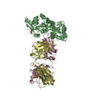
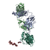
Title
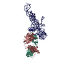
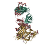
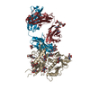
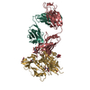
Crystal structure of maltose bound MBP with a conformationally specific synthetic antigen binder (sAB)
Components
  • Maltose-binding periplasmic protein
  • SAB Heavy Chain
  • SAB Light Chain
KeywordsMALTODEXTRIN BINDING PROTEIN/DE NOVO PROTEIN / Maltodextrin binding protein / FAB / antibody fragment / engineered binding protein / MALTODEXTRIN BINDING PROTEIN-DE NOVO PROTEIN complex
Function / homology
Function and homology information


detection of maltose stimulus / maltose binding / maltose transport complex / maltose transport / maltodextrin transmembrane transport / immunoglobulin complex / carbohydrate transmembrane transporter activity / ATP-binding cassette (ABC) transporter complex, substrate-binding subunit-containing / carbohydrate transport / ATP-binding cassette (ABC) transporter complex ...detection of maltose stimulus / maltose binding / maltose transport complex / maltose transport / maltodextrin transmembrane transport / immunoglobulin complex / carbohydrate transmembrane transporter activity / ATP-binding cassette (ABC) transporter complex, substrate-binding subunit-containing / carbohydrate transport / ATP-binding cassette (ABC) transporter complex / cell chemotaxis / outer membrane-bounded periplasmic space / adaptive immune response / periplasmic space / DNA damage response / extracellular region / membrane / plasma membrane
Similarity search - Function
Maltose/Cyclodextrin ABC transporter, substrate-binding protein / Solute-binding family 1, conserved site / Bacterial extracellular solute-binding proteins, family 1 signature. / Bacterial extracellular solute-binding protein / Bacterial extracellular solute-binding protein / Periplasmic binding protein-like II / Immunoglobulin V-Type / Immunoglobulin V-set domain / D-Maltodextrin-Binding Protein; domain 2 / Immunoglobulin V-set domain ...Maltose/Cyclodextrin ABC transporter, substrate-binding protein / Solute-binding family 1, conserved site / Bacterial extracellular solute-binding proteins, family 1 signature. / Bacterial extracellular solute-binding protein / Bacterial extracellular solute-binding protein / Periplasmic binding protein-like II / Immunoglobulin V-Type / Immunoglobulin V-set domain / D-Maltodextrin-Binding Protein; domain 2 / Immunoglobulin V-set domain / Immunoglobulin subtype / Immunoglobulin / Immunoglobulin/major histocompatibility complex, conserved site / Immunoglobulins and major histocompatibility complex proteins signature. / Immunoglobulin C-Type / Immunoglobulin C1-set / Immunoglobulin C1-set domain / Ig-like domain profile. / Immunoglobulin-like domain / Immunoglobulin-like domain superfamily / Immunoglobulins / Immunoglobulin-like fold / Immunoglobulin-like / Sandwich / 3-Layer(aba) Sandwich / Mainly Beta / Alpha Beta
Similarity search - Domain/homology
alpha-maltose / IMIDAZOLE / Maltose/maltodextrin-binding periplasmic protein / Immunoglobulin gamma-1 heavy chain / Ig-like domain-containing protein
Similarity search - Component
Biological speciesEscherichia coli (E. coli)
Homo sapiens (human)
MethodX-RAY DIFFRACTION / SYNCHROTRON / MOLECULAR REPLACEMENT / molecular replacement / Resolution: 2.1 Å
AuthorsKossiakoff, A.A. / Duguid, E.M. / Sandstrom, A.
CitationJournal: Nat.Struct.Mol.Biol. / Year: 2011
Title: Allosteric control of ligand-binding affinity using engineered conformation-specific effector proteins.
Authors: Rizk, S.S. / Paduch, M. / Heithaus, J.H. / Duguid, E.M. / Sandstrom, A. / Kossiakoff, A.A.
History
DepositionNov 1, 2010Deposition site: RCSB / Processing site: RCSB
Revision 1.0Mar 9, 2011Provider: repository / Type: Initial release
Revision 1.1Jul 13, 2011Group: Version format compliance
Revision 1.2May 31, 2017Group: Database references
Revision 1.3Nov 8, 2017Group: Refinement description / Category: software
Revision 2.0Jul 29, 2020Group: Atomic model / Data collection ...Atomic model / Data collection / Derived calculations / Structure summary
Category: atom_site / atom_site_anisotrop ...atom_site / atom_site_anisotrop / chem_comp / entity / entity_name_com / pdbx_branch_scheme / pdbx_chem_comp_identifier / pdbx_entity_branch / pdbx_entity_branch_descriptor / pdbx_entity_branch_link / pdbx_entity_branch_list / pdbx_entity_nonpoly / pdbx_molecule_features / pdbx_nonpoly_scheme / pdbx_struct_assembly_gen / pdbx_struct_special_symmetry / struct_asym / struct_conn / struct_site / struct_site_gen
Item: _atom_site.auth_asym_id / _atom_site.auth_seq_id ..._atom_site.auth_asym_id / _atom_site.auth_seq_id / _atom_site.label_asym_id / _atom_site_anisotrop.pdbx_label_asym_id / _chem_comp.name / _chem_comp.type / _entity.formula_weight / _entity.pdbx_description / _entity.pdbx_number_of_molecules / _entity.type / _pdbx_struct_assembly_gen.asym_id_list / _struct_conn.pdbx_leaving_atom_flag / _struct_conn.ptnr1_auth_asym_id / _struct_conn.ptnr1_auth_seq_id / _struct_conn.ptnr2_auth_asym_id / _struct_conn.ptnr2_auth_seq_id / _struct_conn.ptnr2_label_asym_id
Description: Carbohydrate remediation / Provider: repository / Type: Remediation
Revision 2.1Sep 6, 2023Group: Data collection / Database references ...Data collection / Database references / Refinement description / Structure summary
Category: chem_comp / chem_comp_atom ...chem_comp / chem_comp_atom / chem_comp_bond / database_2 / pdbx_initial_refinement_model
Item: _chem_comp.pdbx_synonyms / _database_2.pdbx_DOI / _database_2.pdbx_database_accession

-
Structure visualization

Structure viewerMolecule:
MolmilJmol/JSmol

Downloads & links

-
Assembly

Deposited unit
A: Maltose-binding periplasmic protein
H: SAB Heavy Chain
L: SAB Light Chain
hetero molecules


Theoretical massNumber of molelcules
Total (without water)92,8418
Polymers92,1763
Non-polymers6655
Water7,206400
1


  • Idetical with deposited unit
  • defined by author&software
TypeNameSymmetry operationNumber
identity operation1_555x,y,z1
Buried area7730 Å2
ΔGint-27 kcal/mol
Surface area32660 Å2
MethodPISA
Unit cell
Length a, b, c (Å)63.870, 228.460, 135.570
Angle α, β, γ (deg.)90.00, 90.00, 90.00
Int Tables number20
Space group name H-MC2221
Components on special symmetry positions
IDModelComponents
11H-230-

HOH

-
Components

-
Antibody , 2 types, 2 molecules HL

#2: Antibody SAB Heavy Chain / Immunoglobulin gamma-1 heavy chain NIE


Mass: 24669.441 Da / Num. of mol.: 1
Source method: isolated from a genetically manipulated source
Source: (gene. exp.) Homo sapiens (human) / Gene: DKFZp686P15220, ighg / Plasmid: pfab007 / Production host: Escherichia coli (E. coli) / Strain (production host): actt 55244 (derived from w3110) / References: UniProt: P0DOX5
#3: Antibody SAB Light Chain


Mass: 23330.891 Da / Num. of mol.: 1
Source method: isolated from a genetically manipulated source
Source: (gene. exp.) Homo sapiens (human) / Gene: igk, IGK@ / Plasmid: pfab007 / Production host: Escherichia coli (E. coli) / Strain (production host): actt 55244 (derived from w3110) / References: UniProt: Q8TCD0

-
Protein / Sugars , 2 types, 2 molecules A

#1: Protein Maltose-binding periplasmic protein / MBP / MMBP / Maltodextrin-binding protein


Mass: 44175.613 Da / Num. of mol.: 1 / Mutation: r367n delta (368-370)
Source method: isolated from a genetically manipulated source
Source: (gene. exp.) Escherichia coli (E. coli) / Strain: K12 / Gene: malE, b4034, JW3994 / Plasmid: phft2 / Production host: Escherichia coli (E. coli) / Strain (production host): BL21 / References: UniProt: P0AEX9
#4: Polysaccharide alpha-D-glucopyranose-(1-4)-alpha-D-glucopyranose / alpha-maltose


Type: oligosaccharide, Oligosaccharide / Class: Nutrient / Mass: 342.297 Da / Num. of mol.: 1
Source method: isolated from a genetically manipulated source
Details: oligosaccharide / References: alpha-maltose
DescriptorTypeProgram
DGlcpa1-4DGlcpa1-ROHGlycam Condensed SequenceGMML 1.0
WURCS=2.0/1,2,1/[a2122h-1a_1-5]/1-1/a4-b1WURCSPDB2Glycan 1.1.0
[][a-D-Glcp]{[(4+1)][a-D-Glcp]{}}LINUCSPDB-CARE

-
Non-polymers , 3 types, 404 molecules

#5: Chemical ChemComp-IMD / IMIDAZOLE / Imidazole


Mass: 69.085 Da / Num. of mol.: 2 / Source method: obtained synthetically / Formula: C3H5N2
#6: Chemical ChemComp-GOL / GLYCEROL / GLYCERIN / PROPANE-1,2,3-TRIOL / Glycerol


Mass: 92.094 Da / Num. of mol.: 2 / Source method: obtained synthetically / Formula: C3H8O3
#7: Water ChemComp-HOH / water / Water


Mass: 18.015 Da / Num. of mol.: 400 / Source method: isolated from a natural source / Formula: H2O

-
Details

Sequence detailsTHE SAB HEAVY AND LIGHT CHAINS CONTAIN BOTH A VARIABLE DOMAIN REPRESENTED BY THE SEQUENCE SELF ...THE SAB HEAVY AND LIGHT CHAINS CONTAIN BOTH A VARIABLE DOMAIN REPRESENTED BY THE SEQUENCE SELF REFERENCE AND A CONSTANT DOMAIN THAT REFERENCES THE UNIPROT DATABASE

-
Experimental details

-
Experiment

ExperimentMethod: X-RAY DIFFRACTION / Number of used crystals: 1

-
Sample preparation

CrystalDensity Matthews: 2.68 Å3/Da / Density % sol: 54.15 %
Crystal growTemperature: 292 K / Method: vapor diffusion, hanging drop / pH: 6
Details: 19% PEG 3400, 8% Tacsimate, pH 6.0, VAPOR DIFFUSION, HANGING DROP, temperature 292K

-
Data collection

DiffractionMean temperature: 200 K
Diffraction sourceSource: SYNCHROTRON / Site: APS / Beamline: 21-ID-D / Wavelength: 1.07817 Å
DetectorType: MARMOSAIC 300 mm CCD / Detector: CCD / Date: Jun 3, 2010
RadiationMonochromator: Si(111) / Protocol: SINGLE WAVELENGTH / Monochromatic (M) / Laue (L): M / Scattering type: x-ray
Radiation wavelengthWavelength: 1.07817 Å / Relative weight: 1
ReflectionResolution: 1.83→50 Å / Num. all: 78510 / Num. obs: 74893 / % possible obs: 85.2 % / Observed criterion σ(F): -3 / Redundancy: 4.5 % / Rmerge(I) obs: 0.06 / Net I/σ(I): 12.2
Reflection shell
Resolution (Å)Redundancy (%)Rmerge(I) obs% possible all
1.83-1.862.30.51639.7
1.86-1.92.90.55152.1
1.9-1.933.40.5259.7
1.93-1.973.90.4365.1
1.97-2.014.20.36169
2.01-2.064.30.30372.9
2.06-2.114.40.27178.1
2.11-2.174.40.21984.1
2.17-2.234.40.19989.8
2.23-2.314.50.17795.2
2.31-2.394.60.16198.6
2.39-2.484.80.139100
2.48-2.650.117100
2.6-2.7350.088100
2.73-2.950.07399.9
2.9-3.1350.057100
3.13-3.444.90.05599.7
3.44-3.944.90.04399.6
3.94-4.974.90.03299.4
4.97-504.70.02998.3

-
Phasing

PhasingMethod: molecular replacement
Phasing MRRfactor: 46.6 / Model details: Phaser MODE: MR_AUTO
Highest resolutionLowest resolution
Rotation2.5 Å39.68 Å
Translation2.5 Å39.68 Å

-
Processing

Software
NameVersionClassificationNB
DENZOdata reduction
SCALEPACKdata scaling
PHASER2.1.4phasing
REFMACrefinement
PDB_EXTRACT3.1data extraction
MAR345data collection
RefinementMethod to determine structure: MOLECULAR REPLACEMENT
Starting model: PDB ENTRIES 1ANF and 2R8S
Resolution: 2.1→38.07 Å / Cor.coef. Fo:Fc: 0.949 / Cor.coef. Fo:Fc free: 0.916 / Occupancy max: 1 / Occupancy min: 0.3 / SU B: 9.785 / SU ML: 0.119 / SU R Cruickshank DPI: 0.1912 / Cross valid method: THROUGHOUT / ESU R: 0.187 / ESU R Free: 0.171 / Stereochemistry target values: MAXIMUM LIKELIHOOD / Details: HYDROGENS HAVE BEEN ADDED IN THE RIDING POSITIONS
RfactorNum. reflection% reflectionSelection details
Rfree0.22664 2815 5 %RANDOM
Rwork0.17659 ---
obs0.17903 53794 97.04 %-
all-56609 --
Solvent computationIon probe radii: 0.8 Å / Shrinkage radii: 0.8 Å / VDW probe radii: 1.4 Å / Solvent model: MASK
Displacement parametersBiso mean: 23.964 Å2
Baniso -1Baniso -2Baniso -3
1--0.75 Å20 Å20 Å2
2--0.45 Å20 Å2
3---0.3 Å2
Refinement stepCycle: LAST / Resolution: 2.1→38.07 Å
ProteinNucleic acidLigandSolventTotal
Num. atoms6075 0 45 400 6520
Refine LS restraints
Refine-IDTypeDev idealDev ideal targetNumber
X-RAY DIFFRACTIONr_bond_refined_d0.0250.0226366
X-RAY DIFFRACTIONr_bond_other_d
X-RAY DIFFRACTIONr_angle_refined_deg1.9541.9638687
X-RAY DIFFRACTIONr_angle_other_deg
X-RAY DIFFRACTIONr_dihedral_angle_1_deg13.8955824
X-RAY DIFFRACTIONr_dihedral_angle_2_deg34.90925.08250
X-RAY DIFFRACTIONr_dihedral_angle_3_deg15.985151039
X-RAY DIFFRACTIONr_dihedral_angle_4_deg22.4591517
X-RAY DIFFRACTIONr_chiral_restr0.140.2972
X-RAY DIFFRACTIONr_gen_planes_refined0.0110.0214785
X-RAY DIFFRACTIONr_gen_planes_other
X-RAY DIFFRACTIONr_nbd_refined
X-RAY DIFFRACTIONr_nbd_other
X-RAY DIFFRACTIONr_nbtor_refined
X-RAY DIFFRACTIONr_nbtor_other
X-RAY DIFFRACTIONr_xyhbond_nbd_refined
X-RAY DIFFRACTIONr_xyhbond_nbd_other
X-RAY DIFFRACTIONr_metal_ion_refined
X-RAY DIFFRACTIONr_metal_ion_other
X-RAY DIFFRACTIONr_symmetry_vdw_refined
X-RAY DIFFRACTIONr_symmetry_vdw_other
X-RAY DIFFRACTIONr_symmetry_hbond_refined
X-RAY DIFFRACTIONr_symmetry_hbond_other
X-RAY DIFFRACTIONr_symmetry_metal_ion_refined
X-RAY DIFFRACTIONr_symmetry_metal_ion_other
X-RAY DIFFRACTIONr_mcbond_it0.8421.54006
X-RAY DIFFRACTIONr_mcbond_other
X-RAY DIFFRACTIONr_mcangle_it1.38626481
X-RAY DIFFRACTIONr_scbond_it2.48932356
X-RAY DIFFRACTIONr_scangle_it3.6644.52181
X-RAY DIFFRACTIONr_rigid_bond_restr
X-RAY DIFFRACTIONr_sphericity_free
X-RAY DIFFRACTIONr_sphericity_bonded
LS refinement shellResolution: 2.1→2.154 Å / Total num. of bins used: 20
RfactorNum. reflection% reflection
Rfree0.273 194 -
Rwork0.214 3333 -
obs--82.79 %
Refinement TLS params.

Method: refined / Refine-ID: X-RAY DIFFRACTION

IDL112)L122)L132)L222)L232)L332)S11 (Å °)S12 (Å °)S13 (Å °)S21 (Å °)S22 (Å °)S23 (Å °)S31 (Å °)S32 (Å °)S33 (Å °)T112)T122)T132)T222)T232)T332)Origin x (Å)Origin y (Å)Origin z (Å)
13.1124-0.7432-0.0092.97390.44473.0701-0.02090.1023-0.1358-0.3866-0.10730.40280.008-0.38410.12830.1512-0.0475-0.03620.0786-0.02270.1499-18.2159.881-15.918
20.4297-0.1527-0.09682.00541.171.9305-0.03570.003-0.0363-0.2027-0.00230.0736-0.081-0.03020.0380.0501-0.0160.00410.01230.00070.0325-8.43222.07-2.591
31.2270.0757-0.50122.21670.43042.5591-0.0365-0.0404-0.0871-0.08610.0286-0.16640.08190.13820.00790.0205-0.01290.02740.0303-0.02350.0442-2.55119.0512.277
44.06491.5132-2.54211.4036-1.30544.7833-0.0815-0.6327-0.0861-0.02270.018-0.25950.23290.42780.06350.3320.01770.22610.2031-0.01590.338829.34830.041-16.439
52.88850.5796-1.54690.6136-0.22181.211-0.11970.0037-0.0654-0.26020.064-0.27690.20270.0770.05580.2922-0.05010.19440.0534-0.04160.166618.21929.933-17.368
61.9044-0.145-0.31843.64621.70942.1828-0.232-0.1603-0.0931-0.05460.0132-0.23420.00160.13560.21880.11430.02420.06380.10480.04380.122840.88156.217-12.979
74.506611.23743.844645.85459.33583.9574-0.39140.1226-0.45470.56370.6068-2.369-0.0350.5883-0.21540.3320.22010.0530.41750.03910.392849.37855.112-9.525
81.49790.6061-0.70275.6061-0.41880.8303-0.18460.17410.1389-0.63780.13390.13230.0966-0.11410.05080.1464-0.08310.00950.0859-0.01240.06597.52947.699-17.917
93.6749-0.4022-1.27131.47320.07231.4631-0.0713-0.0021-0.0614-0.0509-0.0337-0.22210.04420.17180.10510.0931-0.01410.01770.05170.00840.046637.98466.211-23.911
Refinement TLS group
IDRefine-IDRefine TLS-IDAuth asym-IDAuth seq-ID
1X-RAY DIFFRACTION1A1 - 49
2X-RAY DIFFRACTION2A50 - 306
3X-RAY DIFFRACTION3A307 - 367
4X-RAY DIFFRACTION4H2 - 22
5X-RAY DIFFRACTION5H23 - 114
6X-RAY DIFFRACTION6H115 - 202
7X-RAY DIFFRACTION7H203 - 216
8X-RAY DIFFRACTION8L1 - 106
9X-RAY DIFFRACTION9L107 - 214

+
About Yorodumi

-
News

-
Feb 9, 2022. New format data for meta-information of EMDB entries

New format data for meta-information of EMDB entries

  • Version 3 of the EMDB header file is now the official format.
  • The previous official version 1.9 will be removed from the archive.

Related info.:EMDB header

External links:wwPDB to switch to version 3 of the EMDB data model

-
Aug 12, 2020. Covid-19 info

Covid-19 info

URL: https://pdbj.org/emnavi/covid19.php

New page: Covid-19 featured information page in EM Navigator.

Related info.:Covid-19 info / Mar 5, 2020. Novel coronavirus structure data

+
Mar 5, 2020. Novel coronavirus structure data

Novel coronavirus structure data

Related info.:Yorodumi Speices / Aug 12, 2020. Covid-19 info

External links:COVID-19 featured content - PDBj / Molecule of the Month (242):Coronavirus Proteases

+
Jan 31, 2019. EMDB accession codes are about to change! (news from PDBe EMDB page)

EMDB accession codes are about to change! (news from PDBe EMDB page)

  • The allocation of 4 digits for EMDB accession codes will soon come to an end. Whilst these codes will remain in use, new EMDB accession codes will include an additional digit and will expand incrementally as the available range of codes is exhausted. The current 4-digit format prefixed with “EMD-” (i.e. EMD-XXXX) will advance to a 5-digit format (i.e. EMD-XXXXX), and so on. It is currently estimated that the 4-digit codes will be depleted around Spring 2019, at which point the 5-digit format will come into force.
  • The EM Navigator/Yorodumi systems omit the EMD- prefix.

Related info.:Q: What is EMD? / ID/Accession-code notation in Yorodumi/EM Navigator

External links:EMDB Accession Codes are Changing Soon! / Contact to PDBj

+
Jul 12, 2017. Major update of PDB

Major update of PDB

  • wwPDB released updated PDB data conforming to the new PDBx/mmCIF dictionary.
  • This is a major update changing the version number from 4 to 5, and with Remediation, in which all the entries are updated.
  • In this update, many items about electron microscopy experimental information are reorganized (e.g. em_software).
  • Now, EM Navigator and Yorodumi are based on the updated data.

External links:wwPDB Remediation / Enriched Model Files Conforming to OneDep Data Standards Now Available in the PDB FTP Archive

-
Yorodumi

Thousand views of thousand structures

  • Yorodumi is a browser for structure data from EMDB, PDB, SASBDB, etc.
  • This page is also the successor to EM Navigator detail page, and also detail information page/front-end page for Omokage search.
  • The word "yorodu" (or yorozu) is an old Japanese word meaning "ten thousand". "mi" (miru) is to see.

Related info.:EMDB / PDB / SASBDB / Comparison of 3 databanks / Yorodumi Search / Aug 31, 2016. New EM Navigator & Yorodumi / Yorodumi Papers / Jmol/JSmol / Function and homology information / Changes in new EM Navigator and Yorodumi

Read more