 Movie
Movie Controller

[English] 日本語
 Yorodumi
- PDB-1i8m: CRYSTAL STRUCTURE OF A RECOMBINANT ANTI-SINGLE-STRANDED DNA ANTIB... -
+ Open data

- Basic information
| Entry | Database: PDB / ID: 1i8m | ||||||
|---|---|---|---|---|---|---|---|
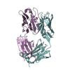
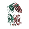
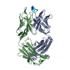
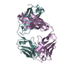
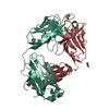
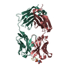
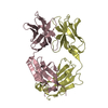
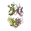
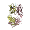
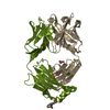
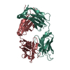
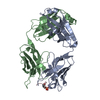
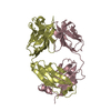
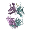
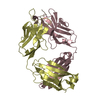
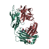
| Title | CRYSTAL STRUCTURE OF A RECOMBINANT ANTI-SINGLE-STRANDED DNA ANTIBODY FRAGMENT COMPLEXED WITH DT5 | ||||||
 Components Components |
| ||||||
 Keywords Keywords | IMMUNE SYSTEM/DNA / fab / antibody / anti-DNA antibody / autoantibody / lupus / IMMUNE SYSTEM-DNA COMPLEX | ||||||
| Function / homology |  Function and homology information Function and homology informationimmunoglobulin complex / adaptive immune response / extracellular region / plasma membrane Similarity search - Function | ||||||
| Biological species |  | ||||||
| Method |  X-RAY DIFFRACTION / SYNCHROTRON / MOLECULAR REPLACEMENT / Resolution: 2.1 Å X-RAY DIFFRACTION / SYNCHROTRON / MOLECULAR REPLACEMENT / Resolution: 2.1 Å | ||||||
 Authors Authors | Tanner, J.J. / Komissarov, A.A. / Deutscher, S.L. | ||||||
 Citation Citation | Journal: J.Mol.Biol. / Year: 2001 Title: Crystal Structure of an Antigen-Binding Fragment Bound to Single-Stranded DNA Authors: Tanner, J.J. / Komissarov, A.A. / Deutscher, S.L. #1: Journal: Acta Crystallogr.,Sect.D / Year: 2000Title: Crystallization and Molecular Replacement Studies of a Recombinant Antigen-Binding Fragment Complexed with Single-stranded DNA. Authors: Prewitt, S.P. / Komissarov, A.A. / Deutscher, S.L. / Tanner, J.J. | ||||||
| History |
|
- Structure visualization
| Structure viewer | Molecule: MolmilJmol/JSmol |
|---|
- Downloads & links
-Download
| PDBx/mmCIF format | 1i8m.cif.gz | 184 KB | Display | PDBx/mmCIF format |
|---|---|---|---|---|
| PDB format | pdb1i8m.ent.gz | 143.8 KB | Display | PDB format |
| PDBx/mmJSON format | 1i8m.json.gz | Tree view | PDBx/mmJSON format | |
| Others |  Other downloads Other downloads |
-Validation report
| Arichive directory | https://data.pdbj.org/pub/pdb/validation_reports/i8/1i8mftp://data.pdbj.org/pub/pdb/validation_reports/i8/1i8m | HTTPS FTP |
|---|
-Related structure data
| Related structure data |  1mlcS S: Starting model for refinement |
|---|---|
| Similar structure data |


-Links
 PDBj
PDBj

- Assembly
| Deposited unit | 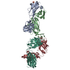
| ||||||||
|---|---|---|---|---|---|---|---|---|---|
| 1 | 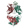
| ||||||||
| 2 | 
| ||||||||
| Unit cell |
| ||||||||
| Details | The biological assembly is an antigen binding fragment, Fab. There are two Fabs in the asymmetric unit. |
-Components
-DNA chain , 2 types, 2 molecules TD
| #1: DNA chain | Mass: 1476.007 Da / Num. of mol.: 1 / Source method: obtained synthetically |
|---|---|
| #2: DNA chain | Mass: 563.428 Da / Num. of mol.: 1 / Source method: obtained synthetically |
-Antibody , 2 types, 4 molecules LAHB
| #3: Antibody | Mass: 23616.057 Da / Num. of mol.: 2 Source method: isolated from a genetically manipulated source Source: (gene. exp.)  #4: Antibody | Mass: 24153.158 Da / Num. of mol.: 2 Source method: isolated from a genetically manipulated source Source: (gene. exp.) |
|---|
-Non-polymers , 2 types, 340 molecules 

| #5: Chemical | ChemComp-SO4 /  Mass: 96.063 Da / Num. of mol.: 1 / Source method: obtained synthetically / Formula: SO4 Mass: 96.063 Da / Num. of mol.: 1 / Source method: obtained synthetically / Formula: SO4 |
|---|---|
| #6: Water | ChemComp-HOH / Mass: 18.015 Da / Num. of mol.: 339 / Source method: isolated from a natural source / Formula: H2O |
-Details
| Has protein modification | Y |
|---|
-Experimental details
-Experiment
| Experiment | Method: X-RAY DIFFRACTION / Number of used crystals: 2 |
|---|
- Sample preparation
| Crystal | Density Matthews: 3.1 Å3/Da / Density % sol: 63 % | ||||||||||||||||||||||||||||||
|---|---|---|---|---|---|---|---|---|---|---|---|---|---|---|---|---|---|---|---|---|---|---|---|---|---|---|---|---|---|---|---|
| Crystal grow | Temperature: 295 K / Method: vapor diffusion, sitting drop / pH: 5 Details: 2 M ammonium sulfate, 0.1 M sodium acetate, pH 5, VAPOR DIFFUSION, SITTING DROP, temperature 295K | ||||||||||||||||||||||||||||||
| Components of the solutions |
| ||||||||||||||||||||||||||||||
| Crystal grow | *PLUS Temperature: 22 ℃ / PH range low: 5.2 / PH range high: 4.8 | ||||||||||||||||||||||||||||||
| Components of the solutions | *PLUS
|
-Data collection
| Diffraction |
| ||||||||||||||||||
|---|---|---|---|---|---|---|---|---|---|---|---|---|---|---|---|---|---|---|---|
| Diffraction source |
| ||||||||||||||||||
| Detector |
| ||||||||||||||||||
| Radiation |
| ||||||||||||||||||
| Radiation wavelength |
| ||||||||||||||||||
| Reflection | Resolution: 2.1→28 Å / Num. all: 72520 / Num. obs: 72520 / % possible obs: 99 % / Observed criterion σ(F): 0 / Observed criterion σ(I): 0 / Redundancy: 6.8 % / Biso Wilson estimate: 23 Å2 / Rmerge(I) obs: 0.079 / Net I/σ(I): 19.3 | ||||||||||||||||||
| Reflection shell | Resolution: 2.1→2.18 Å / Redundancy: 3.6 % / Rmerge(I) obs: 0.294 / Mean I/σ(I) obs: 3.6 / % possible all: 96 | ||||||||||||||||||
| Reflection | *PLUS Lowest resolution: 28 Å / % possible obs: 98.8 % / Num. measured all: 489562 | ||||||||||||||||||
| Reflection shell | *PLUS Highest resolution: 2.1 Å / % possible obs: 95.9 % |

- Processing
| Software |
| ||||||||||||||||||||||||||||||||||||
|---|---|---|---|---|---|---|---|---|---|---|---|---|---|---|---|---|---|---|---|---|---|---|---|---|---|---|---|---|---|---|---|---|---|---|---|---|---|
| Refinement | Method to determine structure: MOLECULAR REPLACEMENT Starting model: 1mlc Resolution: 2.1→27.66 Å / Rfactor Rfree error: 0.003 / Data cutoff high absF: 3972329.88 / Data cutoff low absF: 0 / Isotropic thermal model: RESTRAINED / Cross valid method: THROUGHOUT / σ(F): 0 / σ(I): 0 / Stereochemistry target values: Engh & Huber
| ||||||||||||||||||||||||||||||||||||
| Solvent computation | Solvent model: FLAT MODEL / Bsol: 50.21 Å2 / ksol: 0.383 e/Å3 | ||||||||||||||||||||||||||||||||||||
| Displacement parameters | Biso mean: 38.5 Å2
| ||||||||||||||||||||||||||||||||||||
| Refine analyze |
| ||||||||||||||||||||||||||||||||||||
| Refinement step | Cycle: LAST / Resolution: 2.1→27.66 Å
| ||||||||||||||||||||||||||||||||||||
| Refine LS restraints |
| ||||||||||||||||||||||||||||||||||||
| LS refinement shell | Resolution: 2.1→2.23 Å / Rfactor Rfree error: 0.009 / Total num. of bins used: 6
| ||||||||||||||||||||||||||||||||||||
| Xplor file |
| ||||||||||||||||||||||||||||||||||||
| Software | *PLUS Name: CNS / Version: 1 / Classification: refinement | ||||||||||||||||||||||||||||||||||||
| Refinement | *PLUS σ(F): 0 / Rfactor obs: 0.219 | ||||||||||||||||||||||||||||||||||||
| Solvent computation | *PLUS | ||||||||||||||||||||||||||||||||||||
| Displacement parameters | *PLUS Biso mean: 38.5 Å2 | ||||||||||||||||||||||||||||||||||||
| Refine LS restraints | *PLUS
| ||||||||||||||||||||||||||||||||||||
| LS refinement shell | *PLUS Rfactor Rfree: 0.304 / % reflection Rfree: 9.3 % / Rfactor obs: 0.274 |
