 Movie
Movie Controller

[English] 日本語
 Yorodumi
- PDB-5b3j: Activation of NMDA receptors and the mechanism of inhibition by i... -
+ Open data

- Basic information
| Entry | Database: PDB / ID: 5b3j | |||||||||
|---|---|---|---|---|---|---|---|---|---|---|
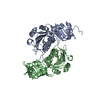
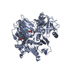
| Title | Activation of NMDA receptors and the mechanism of inhibition by ifenprodil | |||||||||
 Components Components |
| |||||||||
 Keywords Keywords | TRANSPORT PROTEIN / NMDA receptor | |||||||||
| Function / homology |  Function and homology information Function and homology informationcellular response to corticosterone stimulus / cellular response to magnesium starvation / sensory organ development / cellular response to curcumin / regulation of cAMP/PKA signal transduction / EPHB-mediated forward signaling / Assembly and cell surface presentation of NMDA receptors / auditory behavior / sensitization / response to other organism ...cellular response to corticosterone stimulus / cellular response to magnesium starvation / sensory organ development / cellular response to curcumin / regulation of cAMP/PKA signal transduction / EPHB-mediated forward signaling / Assembly and cell surface presentation of NMDA receptors / auditory behavior / sensitization / response to other organism / response to hydrogen sulfide / dendritic branch / fear response / response to methylmercury / regulation of ARF protein signal transduction / apical dendrite / response to manganese ion / suckling behavior / interleukin-1 receptor binding / response to carbohydrate / cellular response to lipid / cellular response to dsRNA / RAF/MAP kinase cascade / negative regulation of dendritic spine maintenance / positive regulation of inhibitory postsynaptic potential / response to amine / response to growth hormone / heterocyclic compound binding / Synaptic adhesion-like molecules / response to glycoside / regulation of monoatomic cation transmembrane transport / NMDA glutamate receptor activity / NMDA selective glutamate receptor complex / glutamate binding / response to zinc ion / ligand-gated sodium channel activity / calcium ion transmembrane import into cytosol / positive regulation of glutamate secretion / protein heterotetramerization / small molecule binding / glycine binding / receptor clustering / startle response / parallel fiber to Purkinje cell synapse / regulation of MAPK cascade / regulation of postsynaptic membrane potential / behavioral response to pain / response to electrical stimulus / monoatomic cation transmembrane transport / extracellularly glutamate-gated ion channel activity / associative learning / action potential / response to magnesium ion / Unblocking of NMDA receptors, glutamate binding and activation / regulation of neuronal synaptic plasticity / monoatomic cation transport / glutamate receptor binding / detection of mechanical stimulus involved in sensory perception of pain / ligand-gated monoatomic ion channel activity / multicellular organismal response to stress / response to mechanical stimulus / neuron development / long-term memory / postsynaptic density, intracellular component / behavioral fear response / monoatomic cation channel activity / synaptic cleft / response to fungicide / cellular response to manganese ion / glutamate-gated receptor activity / regulation of long-term synaptic depression / positive regulation of synaptic transmission, glutamatergic / glutamate-gated calcium ion channel activity / presynaptic active zone membrane / response to cytokine / D2 dopamine receptor binding / cell adhesion molecule binding / ionotropic glutamate receptor signaling pathway / ionotropic glutamate receptor binding / ligand-gated monoatomic ion channel activity involved in regulation of presynaptic membrane potential / protein tyrosine kinase binding / cellular response to forskolin / positive regulation of excitatory postsynaptic potential / hippocampal mossy fiber to CA3 synapse / protein serine/threonine kinase binding / sodium ion transmembrane transport / learning / response to nicotine / synaptic membrane / response to amphetamine / hippocampus development / response to cocaine / cellular response to amino acid stimulus / transmitter-gated monoatomic ion channel activity involved in regulation of postsynaptic membrane potential / synaptic transmission, glutamatergic / regulation of membrane potential / excitatory postsynaptic potential / cerebral cortex development / response to calcium ion / regulation of synaptic plasticity Similarity search - Function | |||||||||
| Biological species |  | |||||||||
| Method |  X-RAY DIFFRACTION / SYNCHROTRON / MOLECULAR REPLACEMENT / Resolution: 2.9 Å X-RAY DIFFRACTION / SYNCHROTRON / MOLECULAR REPLACEMENT / Resolution: 2.9 Å | |||||||||
 Authors Authors | Tajima, N. / Karakas, E. / Grant, T. / Simorowski, N. / Diaz-Avalos, R. / Grigorieff, N. / Furukawa, H. | |||||||||
| Funding support |  United States, 2items United States, 2items
| |||||||||
 Citation Citation | Journal: Nature / Year: 2016 Title: Activation of NMDA receptors and the mechanism of inhibition by ifenprodil. Authors: Nami Tajima / Erkan Karakas / Timothy Grant / Noriko Simorowski / Ruben Diaz-Avalos / Nikolaus Grigorieff / Hiro Furukawa / Abstract: The physiology of N-methyl-d-aspartate (NMDA) receptors is fundamental to brain development and function. NMDA receptors are ionotropic glutamate receptors that function as heterotetramers composed ...The physiology of N-methyl-d-aspartate (NMDA) receptors is fundamental to brain development and function. NMDA receptors are ionotropic glutamate receptors that function as heterotetramers composed mainly of GluN1 and GluN2 subunits. Activation of NMDA receptors requires binding of neurotransmitter agonists to a ligand-binding domain (LBD) and structural rearrangement of an amino-terminal domain (ATD). Recent crystal structures of GluN1-GluN2B NMDA receptors bound to agonists and an allosteric inhibitor, ifenprodil, represent the allosterically inhibited state. However, how the ATD and LBD move to activate the NMDA receptor ion channel remains unclear. Here we applied X-ray crystallography, single-particle electron cryomicroscopy and electrophysiology to rat NMDA receptors to show that, in the absence of ifenprodil, the bi-lobed structure of GluN2 ATD adopts an open conformation accompanied by rearrangement of the GluN1-GluN2 ATD heterodimeric interface, altering subunit orientation in the ATD and LBD and forming an active receptor conformation that gates the ion channel. | |||||||||
| History |
|
- Structure visualization
| Structure viewer | Molecule: MolmilJmol/JSmol |
|---|
- Downloads & links
-Download
| PDBx/mmCIF format | 5b3j.cif.gz | 414.1 KB | Display | PDBx/mmCIF format |
|---|---|---|---|---|
| PDB format | pdb5b3j.ent.gz | 324.2 KB | Display | PDB format |
| PDBx/mmJSON format | 5b3j.json.gz | Tree view | PDBx/mmJSON format | |
| Others |  Other downloads Other downloads |
-Validation report
| Arichive directory | https://data.pdbj.org/pub/pdb/validation_reports/b3/5b3jftp://data.pdbj.org/pub/pdb/validation_reports/b3/5b3j | HTTPS FTP |
|---|
-Related structure data
| Related structure data | 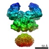 3352C 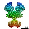 3353C 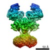 3354C 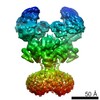 3355C 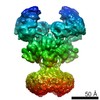 3356C  5fxgC  5fxhC  5fxiC  5fxjC 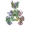 5fxkC C: citing same article ( |
|---|---|
| Similar structure data |


-Links
 PDBj
PDBj


- Assembly
| Deposited unit | 
| ||||||||
|---|---|---|---|---|---|---|---|---|---|
| 1 | 
| ||||||||
| 2 | 
| ||||||||
| 3 | 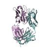
| ||||||||
| 4 | 
| ||||||||
| Unit cell |
|
-Components
-Protein , 2 types, 4 molecules ABCD
| #1: Protein | Mass: 42932.055 Da / Num. of mol.: 2 / Fragment: UNP residues 23-405 / Mutation: N61Q, N371Q Source method: isolated from a genetically manipulated source Source: (gene. exp.)  Trichoplusia ni (cabbage looper) / References: UniProt: Q91977, UniProt: A0A1L8F5J9*PLUS Trichoplusia ni (cabbage looper) / References: UniProt: Q91977, UniProt: A0A1L8F5J9*PLUS#2: Protein | Mass: 41367.902 Da / Num. of mol.: 2 / Fragment: UNP residues 31-394 / Mutation: N348D Source method: isolated from a genetically manipulated source Source: (gene. exp.) Trichoplusia ni (cabbage looper) / References: UniProt: Q00960 |
|---|
-Antibody , 2 types, 4 molecules EHFL
| #3: Antibody | Mass: 23914.783 Da / Num. of mol.: 2 / Source method: isolated from a natural source / Source: (natural) #4: Antibody | Mass: 23675.170 Da / Num. of mol.: 2 / Source method: isolated from a natural source / Source: (natural) |
|---|
-Non-polymers , 2 types, 108 molecules 

| #5: Chemical | ChemComp-NA /  Mass: 22.990 Da / Num. of mol.: 1 / Source method: obtained synthetically / Formula: Na Mass: 22.990 Da / Num. of mol.: 1 / Source method: obtained synthetically / Formula: Na |
|---|---|
| #6: Water | ChemComp-HOH / Mass: 18.015 Da / Num. of mol.: 107 / Source method: isolated from a natural source / Formula: H2O |
-Details
| Has protein modification | Y |
|---|
-Experimental details
-Experiment
| Experiment | Method: X-RAY DIFFRACTION / Number of used crystals: 1 |
|---|
- Sample preparation
| Crystal | Density Matthews: 2.71 Å3/Da / Density % sol: 54.59 % |
|---|---|
| Crystal grow | Temperature: 291 K / Method: vapor diffusion, hanging drop / pH: 4.5 Details: 0.1M sodium acetate, 27% PEG3350, 2.2M sodium formate, 0.05 M calcium chloride |
-Data collection
| Diffraction | Mean temperature: 80 K |
|---|---|
| Diffraction source | Source: SYNCHROTRON / Site: APS / Beamline: 23-ID-D / Wavelength: 1 Å |
| Detector | Type: DECTRIS PILATUS3 6M / Detector: PIXEL / Date: Apr 14, 2014 |
| Radiation | Protocol: SINGLE WAVELENGTH / Monochromatic (M) / Laue (L): M / Scattering type: x-ray |
| Radiation wavelength | Wavelength: 1 Å / Relative weight: 1 |
| Reflection | Resolution: 2.9→50 Å / Num. obs: 57592 / % possible obs: 91.4 % / Redundancy: 4 % / Rmerge(I) obs: 0.099 / Net I/σ(I): 8.5 |
- Processing
| Software |
| ||||||||||||||||||||||||||||||||||||||||||||||||||||||||||||||||||||||||||||||||||||||||||||||||||||||||||||||||||||||||||||||||||||||||||||
|---|---|---|---|---|---|---|---|---|---|---|---|---|---|---|---|---|---|---|---|---|---|---|---|---|---|---|---|---|---|---|---|---|---|---|---|---|---|---|---|---|---|---|---|---|---|---|---|---|---|---|---|---|---|---|---|---|---|---|---|---|---|---|---|---|---|---|---|---|---|---|---|---|---|---|---|---|---|---|---|---|---|---|---|---|---|---|---|---|---|---|---|---|---|---|---|---|---|---|---|---|---|---|---|---|---|---|---|---|---|---|---|---|---|---|---|---|---|---|---|---|---|---|---|---|---|---|---|---|---|---|---|---|---|---|---|---|---|---|---|---|---|
| Refinement | Method to determine structure: MOLECULAR REPLACEMENT / Resolution: 2.9→29.93 Å / SU ML: 0.41 / Cross valid method: NONE / σ(F): 1.36 / Phase error: 31.64 / Stereochemistry target values: ML
| ||||||||||||||||||||||||||||||||||||||||||||||||||||||||||||||||||||||||||||||||||||||||||||||||||||||||||||||||||||||||||||||||||||||||||||
| Solvent computation | Shrinkage radii: 0.9 Å / VDW probe radii: 1.11 Å / Solvent model: FLAT BULK SOLVENT MODEL | ||||||||||||||||||||||||||||||||||||||||||||||||||||||||||||||||||||||||||||||||||||||||||||||||||||||||||||||||||||||||||||||||||||||||||||
| Refinement step | Cycle: LAST / Resolution: 2.9→29.93 Å
| ||||||||||||||||||||||||||||||||||||||||||||||||||||||||||||||||||||||||||||||||||||||||||||||||||||||||||||||||||||||||||||||||||||||||||||
| Refine LS restraints |
| ||||||||||||||||||||||||||||||||||||||||||||||||||||||||||||||||||||||||||||||||||||||||||||||||||||||||||||||||||||||||||||||||||||||||||||
| LS refinement shell |
|
