 Movie
Movie Controller

[English] 日本語
 Yorodumi
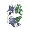
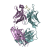
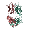
- PDB-2y6s: Structure of an Ebolavirus-protective antibody in complex with it... -
+ Open data

- Basic information
| Entry | Database: PDB / ID: 2y6s | ||||||
|---|---|---|---|---|---|---|---|
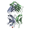
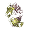
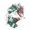
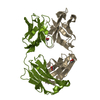
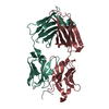
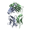
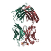
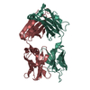
| Title | Structure of an Ebolavirus-protective antibody in complex with its mucin-domain linear epitope | ||||||
 Components Components |
| ||||||
 Keywords Keywords | IMMUNE SYSTEM/VIRAL PROTEIN / IMMUNE SYSTEM-VIRAL PROTEIN COMPLEX | ||||||
| Function / homology |  Function and homology information Function and homology informationsymbiont-mediated-mediated suppression of host tetherin activity / clathrin-dependent endocytosis of virus by host cell / entry receptor-mediated virion attachment to host cell / symbiont-mediated suppression of host innate immune response / fusion of virus membrane with host endosome membrane / viral envelope / symbiont entry into host cell / lipid binding / host cell plasma membrane / virion membrane / extracellular region Similarity search - Function | ||||||
| Biological species |    ZAIRE EBOLAVIRUS ZAIRE EBOLAVIRUS | ||||||
| Method |  X-RAY DIFFRACTION / SYNCHROTRON / MOLECULAR REPLACEMENT / Resolution: 2.8 Å X-RAY DIFFRACTION / SYNCHROTRON / MOLECULAR REPLACEMENT / Resolution: 2.8 Å | ||||||
 Authors Authors | Olal, D.O. / Kuehne, A. / Lee, J.E. / Bale, S. / Dye, J.M. / Saphire, E.O. | ||||||
 Citation Citation | Journal: J.Virol. / Year: 2012 Title: Structure of an Ebola Virus-Protective Antibody in Complex with its Mucin-Domain Linear Epitope. Authors: Olal, D.O. / Kuehne, A. / Bale, S. / Halfmann, P. / Hashiguchi, T. / Fusco, M.L. / Lee, J.E. / King, L.B. / Kawaoka, Y. / Dye, J.M. / Saphire, E.O. | ||||||
| History |
|
- Structure visualization
| Structure viewer | Molecule: MolmilJmol/JSmol |
|---|
- Downloads & links
-Download
| PDBx/mmCIF format | 2y6s.cif.gz | 175.8 KB | Display | PDBx/mmCIF format |
|---|---|---|---|---|
| PDB format | pdb2y6s.ent.gz | 140 KB | Display | PDB format |
| PDBx/mmJSON format | 2y6s.json.gz | Tree view | PDBx/mmJSON format | |
| Others |  Other downloads Other downloads |
-Validation report
| Arichive directory | https://data.pdbj.org/pub/pdb/validation_reports/y6/2y6sftp://data.pdbj.org/pub/pdb/validation_reports/y6/2y6s | HTTPS FTP |
|---|
-Related structure data
| Related structure data | 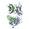 3dggS S: Starting model for refinement |
|---|---|
| Similar structure data |


-Links
 PDBj
PDBj


- Assembly
| Deposited unit | 
| ||||||||||||||||||||||||||||||||||||||||||||||||||||||||||||||
|---|---|---|---|---|---|---|---|---|---|---|---|---|---|---|---|---|---|---|---|---|---|---|---|---|---|---|---|---|---|---|---|---|---|---|---|---|---|---|---|---|---|---|---|---|---|---|---|---|---|---|---|---|---|---|---|---|---|---|---|---|---|---|---|
| 1 | 
| ||||||||||||||||||||||||||||||||||||||||||||||||||||||||||||||
| 2 | 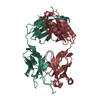
| ||||||||||||||||||||||||||||||||||||||||||||||||||||||||||||||
| Unit cell |
| ||||||||||||||||||||||||||||||||||||||||||||||||||||||||||||||
| Noncrystallographic symmetry (NCS) | NCS domain:
NCS domain segments:
NCS ensembles :
NCS oper:
|
-Components
| #1: Antibody | Mass: 23762.254 Da / Num. of mol.: 2 / Source method: isolated from a natural source / Details: HYBRIDOMA / Source: (natural) #2: Antibody | Mass: 22797.408 Da / Num. of mol.: 2 / Source method: isolated from a natural source / Details: HYBRIDOMA / Source: (natural) #3: Protein/peptide | Mass: 1611.946 Da / Num. of mol.: 2 / Fragment: RESIDUES 477-493 / Source method: obtained synthetically / Source: (synth.) ZAIRE EBOLAVIRUS / References: UniProt: P87666#4: Water | ChemComp-HOH / |  Mass: 18.015 Da / Num. of mol.: 48 / Source method: isolated from a natural source / Formula: H2O Mass: 18.015 Da / Num. of mol.: 48 / Source method: isolated from a natural source / Formula: H2OHas protein modification | Y | |
|---|
-Experimental details
-Experiment
| Experiment | Method: X-RAY DIFFRACTION / Number of used crystals: 1 |
|---|
- Sample preparation
| Crystal | Density Matthews: 2.62 Å3/Da / Density % sol: 53.1 % / Description: NONE |
|---|---|
| Crystal grow | pH: 6.5 / Details: 0.1M BIS-TRIS PROPANE PH6.5, 20% PEG 3350 |
-Data collection
| Diffraction | Mean temperature: 100 K | |||||||||
|---|---|---|---|---|---|---|---|---|---|---|
| Diffraction source | Source: SYNCHROTRON / Site: SSRL  / Beamline: BL11-1 / Wavelength: 0.82, 1.17 / Beamline: BL11-1 / Wavelength: 0.82, 1.17 | |||||||||
| Detector | Type: MARMOSAIC 325 mm CCD / Detector: CCD / Date: May 13, 2010 / Details: MIRRORS | |||||||||
| Radiation | Monochromator: SIDE SCATTERING BENT CUBE ROOT I BEAM SINGLE CRYSTAL, ASYMMETRIC CUT 4.965 DEGS Protocol: SINGLE WAVELENGTH / Monochromatic (M) / Laue (L): M / Scattering type: x-ray | |||||||||
| Radiation wavelength |
| |||||||||
| Reflection | Resolution: 2.3→50 Å / Num. obs: 46342 / % possible obs: 93.9 % / Observed criterion σ(I): 2 / Redundancy: 1.7 % / Biso Wilson estimate: 44.75 Å2 / Rmerge(I) obs: 0.04 / Net I/σ(I): 19.9 | |||||||||
| Reflection shell | Resolution: 2.8→50 Å / Redundancy: 1.7 % / Rmerge(I) obs: 0.2 / Mean I/σ(I) obs: 2.2 / % possible all: 85.1 |
- Processing
| Software |
| ||||||||||||||||||||||||||||||||||||||||||||||||||||||||||||||||||||||
|---|---|---|---|---|---|---|---|---|---|---|---|---|---|---|---|---|---|---|---|---|---|---|---|---|---|---|---|---|---|---|---|---|---|---|---|---|---|---|---|---|---|---|---|---|---|---|---|---|---|---|---|---|---|---|---|---|---|---|---|---|---|---|---|---|---|---|---|---|---|---|---|
| Refinement | Method to determine structure: MOLECULAR REPLACEMENT Starting model: PDB ENTRY 3DGG Resolution: 2.8→38.122 Å / SU ML: 0.41 / σ(F): 0 / Phase error: 31.64 / Stereochemistry target values: ML / Details: RESIDUES 127-133 ARE DISORDERED
| ||||||||||||||||||||||||||||||||||||||||||||||||||||||||||||||||||||||
| Solvent computation | Shrinkage radii: 0.9 Å / VDW probe radii: 1.11 Å / Solvent model: FLAT BULK SOLVENT MODEL / Bsol: 26.94 Å2 / ksol: 0.334 e/Å3 | ||||||||||||||||||||||||||||||||||||||||||||||||||||||||||||||||||||||
| Displacement parameters | Biso mean: 45.6 Å2
| ||||||||||||||||||||||||||||||||||||||||||||||||||||||||||||||||||||||
| Refinement step | Cycle: LAST / Resolution: 2.8→38.122 Å
| ||||||||||||||||||||||||||||||||||||||||||||||||||||||||||||||||||||||
| Refine LS restraints |
| ||||||||||||||||||||||||||||||||||||||||||||||||||||||||||||||||||||||
| Refine LS restraints NCS |
| ||||||||||||||||||||||||||||||||||||||||||||||||||||||||||||||||||||||
| LS refinement shell |
|
