 Movie
Movie Controller

[English] 日本語
 Yorodumi
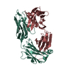
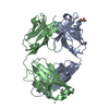
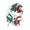
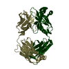
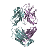
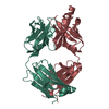
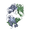
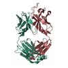
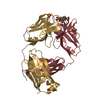
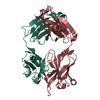
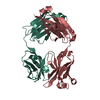
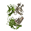
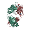
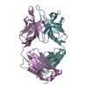
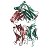
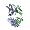
- PDB-1it9: CRYSTAL STRUCTURE OF AN ANTIGEN-BINDING FRAGMENT FROM A HUMANIZED... -
+ Open data

- Basic information
| Entry | Database: PDB / ID: 1it9 | ||||||
|---|---|---|---|---|---|---|---|
| Title | CRYSTAL STRUCTURE OF AN ANTIGEN-BINDING FRAGMENT FROM A HUMANIZED VERSION OF THE ANTI-HUMAN FAS ANTIBODY HFE7A | ||||||
 Components Components |
| ||||||
 Keywords Keywords | IMMUNE SYSTEM / IMMUNOGLOBULIN / FAB / ANTI_FAS / AGONISTIC_ANTIBODY / APOPTOSIS / ANTIBODY_HUMANIZATION | ||||||
| Function / homology | Immunoglobulins / Immunoglobulin-like / Sandwich / Mainly Beta / : / :  Function and homology information Function and homology information | ||||||
| Biological species |  | ||||||
| Method |  X-RAY DIFFRACTION / SYNCHROTRON / MOLECULAR REPLACEMENT / Resolution: 2.8 Å X-RAY DIFFRACTION / SYNCHROTRON / MOLECULAR REPLACEMENT / Resolution: 2.8 Å | ||||||
 Authors Authors | Ito, S. / Takayama, T. / Hanzawa, H. / Takahashi, T. / Miyadai, K. / Serizawa, N. / Hata, T. / Haruyama, H. | ||||||
 Citation Citation | Journal: BIOL.PHARM.BULL. / Year: 2002 Title: Humanization of the Mouse Anti-Fas Antibody HFE7A and Crystal Structure of the Humanized HFE7A Fab Fragment Authors: Haruyama, H. / Ito, S. / Miyadai, K. / Takahashi, T. / Kawaida, R. / Takayama, T. / Hanzawa, H. / Hata, T. / Yamaguchi, J. / Yoshida-Kato, H. / Ichikawa, K. / Ohsumi, J. / Yonehara, S. / Serizawa, N. #1: Journal: To be PublishedTitle: CRYSTALLIZATION AND PRELIMINARY X-RAY STUDIES OF THE FAB FRAGMENT FROM A HUMANIZED VERSION OF THE MOUSE ANTI-HUMAN FAS ANTIBODY HFE7A Authors: Ito, S. / Takayama, T. / Hanzawa, H. / Takahashi, T. / Miyadai, K. / Serizawa, N. / Haruyama, H. / Hata, T. #2: Journal: J.BIOCHEM.(TOKYO) / Year: 2002Title: Crystal Structure of the Antigen-Binding Fragment of Apoptosis-Inducing Mouse Anti-Human Fas Monoclonal Antibody HFE7A Authors: Ito, S. / Takayama, T. / Hanzawa, H. / Ichikawa, K. / Ohsumi, J. / Serizawa, N. / Hata, T. / Haruyama, H. | ||||||
| History |
|
- Structure visualization
| Structure viewer | Molecule: MolmilJmol/JSmol |
|---|
- Downloads & links
-Download
| PDBx/mmCIF format | 1it9.cif.gz | 94.7 KB | Display | PDBx/mmCIF format |
|---|---|---|---|---|
| PDB format | pdb1it9.ent.gz | 72.4 KB | Display | PDB format |
| PDBx/mmJSON format | 1it9.json.gz | Tree view | PDBx/mmJSON format | |
| Others |  Other downloads Other downloads |
-Validation report
| Arichive directory | https://data.pdbj.org/pub/pdb/validation_reports/it/1it9ftp://data.pdbj.org/pub/pdb/validation_reports/it/1it9 | HTTPS FTP |
|---|
-Related structure data
| Related structure data | |
|---|---|
| Similar structure data |


-Links
 PDBj
PDBj

- Assembly
| Deposited unit | 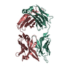
| ||||||||
|---|---|---|---|---|---|---|---|---|---|
| 1 |
| ||||||||
| Unit cell |
|
-Components
| #1: Antibody | Mass: 23501.871 Da / Num. of mol.: 1 / Fragment: FAB FRAGMENT Source method: isolated from a genetically manipulated source Source: (gene. exp.) Chlorocebus aethiops (grivet monkey) / References: UniProt: Q6GMV9 |
|---|---|
| #2: Antibody | Mass: 23593.266 Da / Num. of mol.: 1 / Fragment: FAB FRAGMENT Source method: isolated from a genetically manipulated source Source: (gene. exp.) Chlorocebus aethiops (grivet monkey) / References: UniProt: Q6PJA4 |
| #3: Water | ChemComp-HOH /  Mass: 18.015 Da / Num. of mol.: 6 / Source method: isolated from a natural source / Formula: H2O Mass: 18.015 Da / Num. of mol.: 6 / Source method: isolated from a natural source / Formula: H2O |
| Has protein modification | Y |
-Experimental details
-Experiment
| Experiment | Method: X-RAY DIFFRACTION / Number of used crystals: 2 |
|---|
- Sample preparation
| Crystal | Density Matthews: 2.6 Å3/Da / Density % sol: 51 % | ||||||||||||||||||||||||
|---|---|---|---|---|---|---|---|---|---|---|---|---|---|---|---|---|---|---|---|---|---|---|---|---|---|
| Crystal grow | Temperature: 296 K / Method: vapor diffusion, hanging drop / pH: 4.6 Details: PEG4000, SODIUM ACETATE, METHANOL, pH 4.6, VAPOR DIFFUSION, HANGING DROP, temperature 296K | ||||||||||||||||||||||||
| Crystal grow | *PLUS Temperature: 22 ℃ | ||||||||||||||||||||||||
| Components of the solutions | *PLUS
|
-Data collection
| Diffraction | Mean temperature: 296 K |
|---|---|
| Diffraction source | Source: SYNCHROTRON / Site: Photon Factory  / Beamline: BL-6B / Wavelength: 1 Å / Beamline: BL-6B / Wavelength: 1 Å |
| Detector | Type: WEISSENBERG / Detector: DIFFRACTOMETER / Date: Jun 29, 1999 |
| Radiation | Protocol: SINGLE WAVELENGTH / Monochromatic (M) / Laue (L): M / Scattering type: x-ray |
| Radiation wavelength | Wavelength: 1 Å / Relative weight: 1 |
| Reflection | Resolution: 2.8→50 Å / Num. obs: 12285 / % possible obs: 90.7 % / Observed criterion σ(I): 1 / Redundancy: 6.3 % / Biso Wilson estimate: 42 Å2 / Rmerge(I) obs: 0.082 / Net I/σ(I): 17.1 |
| Reflection shell | Resolution: 2.8→2.9 Å / Rmerge(I) obs: 0.314 / Mean I/σ(I) obs: 5.4 / % possible all: 77.1 |
| Reflection | *PLUS Num. obs: 11136 / % possible obs: 91 % / Num. measured all: 61689 / Rmerge(I) obs: 0.078 |
| Reflection shell | *PLUS Highest resolution: 2.8 Å / Lowest resolution: 2.9 Å / % possible obs: 71.3 % / Num. unique obs: 927 / Num. measured obs: 3790 / Rmerge(I) obs: 0.18 |
- Processing
| Software |
| ||||||||||||||||||||||||||||||||||||||||||||||||||||||||||||||||||||||||||||||||
|---|---|---|---|---|---|---|---|---|---|---|---|---|---|---|---|---|---|---|---|---|---|---|---|---|---|---|---|---|---|---|---|---|---|---|---|---|---|---|---|---|---|---|---|---|---|---|---|---|---|---|---|---|---|---|---|---|---|---|---|---|---|---|---|---|---|---|---|---|---|---|---|---|---|---|---|---|---|---|---|---|---|
| Refinement | Method to determine structure: MOLECULAR REPLACEMENT / Resolution: 2.8→30 Å / Rfactor Rfree error: 0.008 / Data cutoff high absF: 10000000 / Data cutoff low absF: 0.001 / Isotropic thermal model: RESTRAINED / Cross valid method: THROUGHOUT / σ(F): 2 / Stereochemistry target values: Engh & Huber / Details: BULK SOLVENT MODEL USED
| ||||||||||||||||||||||||||||||||||||||||||||||||||||||||||||||||||||||||||||||||
| Displacement parameters |
| ||||||||||||||||||||||||||||||||||||||||||||||||||||||||||||||||||||||||||||||||
| Refine analyze |
| ||||||||||||||||||||||||||||||||||||||||||||||||||||||||||||||||||||||||||||||||
| Refinement step | Cycle: LAST / Resolution: 2.8→30 Å
| ||||||||||||||||||||||||||||||||||||||||||||||||||||||||||||||||||||||||||||||||
| Refine LS restraints |
| ||||||||||||||||||||||||||||||||||||||||||||||||||||||||||||||||||||||||||||||||
| LS refinement shell | Resolution: 2.8→2.98 Å / Rfactor Rfree error: 0.024 / Total num. of bins used: 6
| ||||||||||||||||||||||||||||||||||||||||||||||||||||||||||||||||||||||||||||||||
| Xplor file | Serial no: 1 / Param file: PROTEIN_REP.PARAM / Topol file: TOPHCSDX.TOP | ||||||||||||||||||||||||||||||||||||||||||||||||||||||||||||||||||||||||||||||||
| Refinement | *PLUS Highest resolution: 2.8 Å / Lowest resolution: 30 Å / % reflection Rfree: 9 % / Rfactor Rfree: 0.28 | ||||||||||||||||||||||||||||||||||||||||||||||||||||||||||||||||||||||||||||||||
| Solvent computation | *PLUS | ||||||||||||||||||||||||||||||||||||||||||||||||||||||||||||||||||||||||||||||||
| Displacement parameters | *PLUS | ||||||||||||||||||||||||||||||||||||||||||||||||||||||||||||||||||||||||||||||||
| Refine LS restraints | *PLUS
|
