 Movie
Movie Controller

[English] 日本語
 Yorodumi
- PDB-5gux: Cytochrome c-dependent nitric oxide reductase (cNOR) from Pseudom... -
+ Open data

- Basic information
| Entry | Database: PDB / ID: 5gux | |||||||||
|---|---|---|---|---|---|---|---|---|---|---|
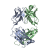
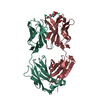
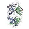
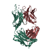
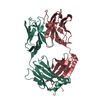
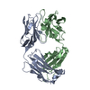
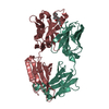
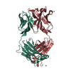
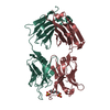
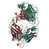
| Title | Cytochrome c-dependent nitric oxide reductase (cNOR) from Pseudomonas aeruginosa in complex with xenon | |||||||||
 Components Components |
| |||||||||
 Keywords Keywords | MEMBRANE PROTEIN / metal-binding | |||||||||
| Function / homology |  Function and homology information Function and homology informationnitric oxide reductase (cytochrome c) / nitric oxide reductase activity / denitrification pathway / cytochrome-c oxidase activity / aerobic respiration / respiratory electron transport chain / electron transfer activity / heme binding / metal ion binding / plasma membrane Similarity search - Function | |||||||||
| Biological species |   Pseudomonas aeruginosa PAO1 (bacteria) Pseudomonas aeruginosa PAO1 (bacteria) | |||||||||
| Method |  X-RAY DIFFRACTION / SYNCHROTRON / MOLECULAR REPLACEMENT / Resolution: 3.3 Å X-RAY DIFFRACTION / SYNCHROTRON / MOLECULAR REPLACEMENT / Resolution: 3.3 Å | |||||||||
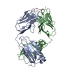
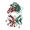
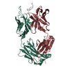
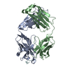
| Model details | nitric oxide reductase and Fab fragment in complex with xenon | |||||||||
 Authors Authors | Ishii, S. / Terasaka, E. / Sugimoto, H. / Shiro, Y. / Tosha, T. | |||||||||
| Funding support |  Japan, 2items Japan, 2items
| |||||||||
 Citation Citation | Journal: Proc. Natl. Acad. Sci. U.S.A. / Year: 2017 Title: Dynamics of nitric oxide controlled by protein complex in bacterial system. Authors: Terasaka, E. / Yamada, K. / Wang, P.H. / Hosokawa, K. / Yamagiwa, R. / Matsumoto, K. / Ishii, S. / Mori, T. / Yagi, K. / Sawai, H. / Arai, H. / Sugimoto, H. / Sugita, Y. / Shiro, Y. / Tosha, T. #1: Journal: Science / Year: 2010Title: Structural basis of biological N2O generation by bacterial nitric oxide reductase. Authors: Hino, T. / Matsumoto, Y. / Nagano, S. / Sugimoto, H. / Fukumori, Y. / Murata, T. / Iwata, S. / Shiro, Y. #2: Journal: Nat. Struct. Mol. Biol. / Year: 2012Title: Crystal structure of quinol-dependent nitric oxide reductase from Geobacillus stearothermophilus. Authors: Matsumoto, Y. / Tosha, T. / Pisliakov, A.V. / Hino, T. / Sugimoto, H. / Nagano, S. / Sugita, Y. / Shiro, Y. #3: Journal: Proteins / Year: 2014Title: Structures of reduced and ligand-bound nitric oxide reductase provide insights into functional differences in respiratory enzymes. Authors: Sato, N. / Ishii, S. / Sugimoto, H. / Hino, T. / Fukumori, Y. / Sako, Y. / Shiro, Y. / Tosha, T. | |||||||||
| History |
|
- Structure visualization
| Structure viewer | Molecule: MolmilJmol/JSmol |
|---|
- Downloads & links
-Download
| PDBx/mmCIF format | 5gux.cif.gz | 426.4 KB | Display | PDBx/mmCIF format |
|---|---|---|---|---|
| PDB format | pdb5gux.ent.gz | 349 KB | Display | PDB format |
| PDBx/mmJSON format | 5gux.json.gz | Tree view | PDBx/mmJSON format | |
| Others |  Other downloads Other downloads |
-Validation report
| Arichive directory | https://data.pdbj.org/pub/pdb/validation_reports/gu/5guxftp://data.pdbj.org/pub/pdb/validation_reports/gu/5gux | HTTPS FTP |
|---|
-Related structure data
| Related structure data |  5guwC  3o0rS S: Starting model for refinement C: citing same article ( |
|---|---|
| Similar structure data |


-Links
 PDBj
PDBj


- Assembly
| Deposited unit | 
| ||||||||
|---|---|---|---|---|---|---|---|---|---|
| 1 | 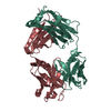
| ||||||||
| 2 | 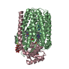
| ||||||||
| Unit cell |
|
-Components
-Nitric oxide reductase subunit ... , 2 types, 2 molecules BC
| #3: Protein | Mass: 52215.871 Da / Num. of mol.: 1 / Fragment: Nitric oxide reductase subunit B / Source method: isolated from a natural source / Details: ? / Source: (natural) Pseudomonas aeruginosa PAO1 (bacteria) / Strain: PAO1References: UniProt: Q59647, nitric oxide reductase (cytochrome c) |
|---|---|
| #4: Protein | Mass: 16374.622 Da / Num. of mol.: 1 Fragment: UNP residues 5-146, Nitric oxide reductase subunit C Mutation: K100N / Source method: isolated from a natural source / Source: (natural) Pseudomonas aeruginosa PAO1 (bacteria) / Strain: PAO1 / References: UniProt: Q59646 |
-Antibody , 2 types, 2 molecules LH
| #1: Antibody | Mass: 23735.326 Da / Num. of mol.: 1 / Fragment: antibody fab fragment light chain / Source method: isolated from a natural source / Source: (natural) |
|---|---|
| #2: Antibody | Mass: 24089.945 Da / Num. of mol.: 1 / Fragment: antibody fab fragment heavy chain / Source method: isolated from a natural source / Source: (natural) |
-Sugars , 1 types, 2 molecules 
| #9: Sugar |  Type: D-saccharide / Mass: 498.628 Da / Num. of mol.: 2 Type: D-saccharide / Mass: 498.628 Da / Num. of mol.: 2Source method: isolated from a genetically manipulated source Formula: C22H42O10S / Comment: detergent*YM |
|---|
-Non-polymers , 6 types, 13 molecules 


| #5: Chemical |  Mass: 616.487 Da / Num. of mol.: 2 / Source method: obtained synthetically / Formula: C34H32FeN4O4 Mass: 616.487 Da / Num. of mol.: 2 / Source method: obtained synthetically / Formula: C34H32FeN4O4#6: Chemical | ChemComp-FE / |  Mass: 55.845 Da / Num. of mol.: 1 / Source method: obtained synthetically / Formula: Fe Mass: 55.845 Da / Num. of mol.: 1 / Source method: obtained synthetically / Formula: Fe#7: Chemical | ChemComp-O / |  Mass: 15.999 Da / Num. of mol.: 1 / Source method: obtained synthetically / Formula: O Mass: 15.999 Da / Num. of mol.: 1 / Source method: obtained synthetically / Formula: O#8: Chemical | ChemComp-XE /  Mass: 131.293 Da / Num. of mol.: 7 / Source method: obtained synthetically / Formula: Xe Mass: 131.293 Da / Num. of mol.: 7 / Source method: obtained synthetically / Formula: Xe#10: Chemical | ChemComp-CA / |  Mass: 40.078 Da / Num. of mol.: 1 / Source method: obtained synthetically / Formula: Ca Mass: 40.078 Da / Num. of mol.: 1 / Source method: obtained synthetically / Formula: Ca#11: Chemical | ChemComp-HEC / |  Mass: 618.503 Da / Num. of mol.: 1 / Source method: obtained synthetically / Formula: C34H34FeN4O4 Mass: 618.503 Da / Num. of mol.: 1 / Source method: obtained synthetically / Formula: C34H34FeN4O4 |
|---|
-Details
| Has protein modification | Y |
|---|
-Experimental details
-Experiment
| Experiment | Method: X-RAY DIFFRACTION / Number of used crystals: 1 |
|---|
- Sample preparation
| Crystal | Density Matthews: 3.99 Å3/Da / Density % sol: 69.15 % |
|---|---|
| Crystal grow | Temperature: 277 K / Method: vapor diffusion / pH: 6 Details: 24% PEG 400, 0.1M SODIUM CITRATE, 50mM SODIUM CHLORIDE, 20mM MAGNESIUM CHLORIDE |
-Data collection
| Diffraction | Mean temperature: 100 K | ||||||||||||||||||||||||||||||||||||||||||||||||||||||||||||||||||||||||||||||||||||||||||||||||||||||||||||||||||||||||||||||
|---|---|---|---|---|---|---|---|---|---|---|---|---|---|---|---|---|---|---|---|---|---|---|---|---|---|---|---|---|---|---|---|---|---|---|---|---|---|---|---|---|---|---|---|---|---|---|---|---|---|---|---|---|---|---|---|---|---|---|---|---|---|---|---|---|---|---|---|---|---|---|---|---|---|---|---|---|---|---|---|---|---|---|---|---|---|---|---|---|---|---|---|---|---|---|---|---|---|---|---|---|---|---|---|---|---|---|---|---|---|---|---|---|---|---|---|---|---|---|---|---|---|---|---|---|---|---|---|
| Diffraction source | Source: SYNCHROTRON / Site: SPring-8 / Beamline: BL41XU / Wavelength: 1 Å | ||||||||||||||||||||||||||||||||||||||||||||||||||||||||||||||||||||||||||||||||||||||||||||||||||||||||||||||||||||||||||||||
| Detector | Type: RAYONIX MX225HE / Detector: CCD / Date: Jun 21, 2013 / Details: mirrors | ||||||||||||||||||||||||||||||||||||||||||||||||||||||||||||||||||||||||||||||||||||||||||||||||||||||||||||||||||||||||||||||
| Radiation | Monochromator: Si(111) / Protocol: SINGLE WAVELENGTH / Monochromatic (M) / Laue (L): M / Scattering type: x-ray | ||||||||||||||||||||||||||||||||||||||||||||||||||||||||||||||||||||||||||||||||||||||||||||||||||||||||||||||||||||||||||||||
| Radiation wavelength | Wavelength: 1 Å / Relative weight: 1 | ||||||||||||||||||||||||||||||||||||||||||||||||||||||||||||||||||||||||||||||||||||||||||||||||||||||||||||||||||||||||||||||
| Reflection | Resolution: 3.3→40 Å / Num. obs: 27449 / % possible obs: 98.3 % / Redundancy: 4.5 % / Rmerge(I) obs: 0.131 / Net I/av σ(I): 10.05 / Net I/σ(I): 7.3 | ||||||||||||||||||||||||||||||||||||||||||||||||||||||||||||||||||||||||||||||||||||||||||||||||||||||||||||||||||||||||||||||
| Reflection shell |
|
- Processing
| Software |
| ||||||||||||||||||||||||||||||||||||||||||||||||||||||||||||||||||||||||||||||||||||||||||||||||||||||||||||||||||||||||||||||||||||||||||||||||||||||||||||||||||||||||||||||||||||||||||||||||||||||||
|---|---|---|---|---|---|---|---|---|---|---|---|---|---|---|---|---|---|---|---|---|---|---|---|---|---|---|---|---|---|---|---|---|---|---|---|---|---|---|---|---|---|---|---|---|---|---|---|---|---|---|---|---|---|---|---|---|---|---|---|---|---|---|---|---|---|---|---|---|---|---|---|---|---|---|---|---|---|---|---|---|---|---|---|---|---|---|---|---|---|---|---|---|---|---|---|---|---|---|---|---|---|---|---|---|---|---|---|---|---|---|---|---|---|---|---|---|---|---|---|---|---|---|---|---|---|---|---|---|---|---|---|---|---|---|---|---|---|---|---|---|---|---|---|---|---|---|---|---|---|---|---|---|---|---|---|---|---|---|---|---|---|---|---|---|---|---|---|---|---|---|---|---|---|---|---|---|---|---|---|---|---|---|---|---|---|---|---|---|---|---|---|---|---|---|---|---|---|---|---|---|---|
| Refinement | Method to determine structure: MOLECULAR REPLACEMENT Starting model: 3O0R Resolution: 3.3→39.39 Å / Cor.coef. Fo:Fc: 0.907 / Cor.coef. Fo:Fc free: 0.866 / SU B: 47.351 / SU ML: 0.358 / SU R Cruickshank DPI: 0.4229 / Cross valid method: THROUGHOUT / σ(F): 0 / ESU R Free: 0.511 Details: HYDROGENS HAVE BEEN USED IF PRESENT IN THE INPUT U VALUES
| ||||||||||||||||||||||||||||||||||||||||||||||||||||||||||||||||||||||||||||||||||||||||||||||||||||||||||||||||||||||||||||||||||||||||||||||||||||||||||||||||||||||||||||||||||||||||||||||||||||||||
| Solvent computation | Ion probe radii: 0.8 Å / Shrinkage radii: 0.8 Å / VDW probe radii: 1.2 Å | ||||||||||||||||||||||||||||||||||||||||||||||||||||||||||||||||||||||||||||||||||||||||||||||||||||||||||||||||||||||||||||||||||||||||||||||||||||||||||||||||||||||||||||||||||||||||||||||||||||||||
| Displacement parameters | Biso max: 222.63 Å2 / Biso mean: 88.715 Å2 / Biso min: 45.1 Å2
| ||||||||||||||||||||||||||||||||||||||||||||||||||||||||||||||||||||||||||||||||||||||||||||||||||||||||||||||||||||||||||||||||||||||||||||||||||||||||||||||||||||||||||||||||||||||||||||||||||||||||
| Refinement step | Cycle: final / Resolution: 3.3→39.39 Å
| ||||||||||||||||||||||||||||||||||||||||||||||||||||||||||||||||||||||||||||||||||||||||||||||||||||||||||||||||||||||||||||||||||||||||||||||||||||||||||||||||||||||||||||||||||||||||||||||||||||||||
| Refine LS restraints |
| ||||||||||||||||||||||||||||||||||||||||||||||||||||||||||||||||||||||||||||||||||||||||||||||||||||||||||||||||||||||||||||||||||||||||||||||||||||||||||||||||||||||||||||||||||||||||||||||||||||||||
| LS refinement shell | Resolution: 3.3→3.385 Å / Total num. of bins used: 20
| ||||||||||||||||||||||||||||||||||||||||||||||||||||||||||||||||||||||||||||||||||||||||||||||||||||||||||||||||||||||||||||||||||||||||||||||||||||||||||||||||||||||||||||||||||||||||||||||||||||||||
| Refinement TLS params. | Method: refined / Refine-ID: X-RAY DIFFRACTION
| ||||||||||||||||||||||||||||||||||||||||||||||||||||||||||||||||||||||||||||||||||||||||||||||||||||||||||||||||||||||||||||||||||||||||||||||||||||||||||||||||||||||||||||||||||||||||||||||||||||||||
| Refinement TLS group |
|
