 Movie
Movie Controller


+ Open data

- Basic information
| Entry | Database: PDB / ID: 3ojd | ||||||
|---|---|---|---|---|---|---|---|
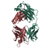
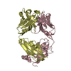
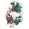
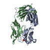
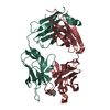
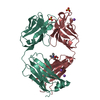
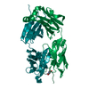
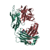
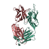
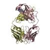
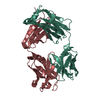
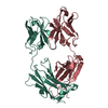
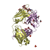
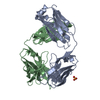
| Title | Anti-Indolicidin monoclonal antibody V2D2 (Fab fragment) | ||||||
 Components Components | (Fab V2D2) x 2 | ||||||
 Keywords Keywords | IMMUNE SYSTEM / Beta-sandwich Immunoglobulin fold / Antigen binding / Secreted protein / anti-indolicidin Fab / surrogate receptor | ||||||
| Function / homology | Immunoglobulins / Immunoglobulin-like / Sandwich / Mainly Beta Function and homology information Function and homology information | ||||||
| Biological species |  | ||||||
| Method |  X-RAY DIFFRACTION / MOLECULAR REPLACEMENT / Resolution: 2 Å X-RAY DIFFRACTION / MOLECULAR REPLACEMENT / Resolution: 2 Å | ||||||
 Authors Authors | Lomash, S. / Salunke, D.M. | ||||||
 Citation Citation | Journal: J.Biol.Chem. / Year: 2010 Title: An antibody as surrogate receptor reveals determinants of activity of an innate immune peptide antibiotic Authors: Lomash, S. / Nagpal, S. / Salunke, D.M. | ||||||
| History |
|
- Structure visualization
| Structure viewer | Molecule: MolmilJmol/JSmol |
|---|
- Downloads & links
-Download
| PDBx/mmCIF format | 3ojd.cif.gz | 105.4 KB | Display | PDBx/mmCIF format |
|---|---|---|---|---|
| PDB format | pdb3ojd.ent.gz | 79.4 KB | Display | PDB format |
| PDBx/mmJSON format | 3ojd.json.gz | Tree view | PDBx/mmJSON format | |
| Others |  Other downloads Other downloads |
-Validation report
| Arichive directory | https://data.pdbj.org/pub/pdb/validation_reports/oj/3ojdftp://data.pdbj.org/pub/pdb/validation_reports/oj/3ojd | HTTPS FTP |
|---|
-Related structure data
| Related structure data |  1ymhS S: Starting model for refinement |
|---|---|
| Similar structure data |


-Links
 PDBj
PDBj

- Assembly
| Deposited unit | 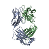
| ||||||||
|---|---|---|---|---|---|---|---|---|---|
| 1 |
| ||||||||
| Unit cell |
|
-Components
| #1: Antibody | Mass: 23415.777 Da / Num. of mol.: 1 / Source method: isolated from a natural source / Details: Light chain / Source: (natural) |
|---|---|
| #2: Antibody | Mass: 24398.447 Da / Num. of mol.: 1 / Source method: isolated from a natural source / Details: Heavy chain / Source: (natural) |
| #3: Water | ChemComp-HOH /  Mass: 18.015 Da / Num. of mol.: 388 / Source method: isolated from a natural source / Formula: H2O Mass: 18.015 Da / Num. of mol.: 388 / Source method: isolated from a natural source / Formula: H2O |
| Has protein modification | Y |
-Experimental details
-Experiment
| Experiment | Method: X-RAY DIFFRACTION / Number of used crystals: 1 |
|---|
- Sample preparation
| Crystal | Density Matthews: 2.25 Å3/Da / Density % sol: 45.28 % |
|---|---|
| Crystal grow | Temperature: 298 K / Method: vapor diffusion, hanging drop / pH: 7.1 Details: 10% PEG 1000, pH 7.1, VAPOR DIFFUSION, HANGING DROP, temperature 298K |
-Data collection
| Diffraction | Mean temperature: 120 K |
|---|---|
| Diffraction source | Source: ROTATING ANODE / Type: RIGAKU RU300 / Wavelength: 1.5418 Å |
| Detector | Type: MARRESEARCH / Detector: IMAGE PLATE / Date: Aug 21, 2006 / Details: mirrors |
| Radiation | Monochromator: Ni FILTER CMF12 38Cu-6 (Osmic Inc.) / Protocol: SINGLE WAVELENGTH / Monochromatic (M) / Laue (L): M / Scattering type: x-ray |
| Radiation wavelength | Wavelength: 1.5418 Å / Relative weight: 1 |
| Reflection | Resolution: 2→50 Å / Num. all: 31660 / Num. obs: 28494 / % possible obs: 90 % / Observed criterion σ(F): 2 / Observed criterion σ(I): 5 / Redundancy: 5.5 % / Rmerge(I) obs: 0.113 / Net I/σ(I): 10.5 |
| Reflection shell | Resolution: 1.96→2.07 Å / Redundancy: 5.9 % / Rmerge(I) obs: 0.526 / Mean I/σ(I) obs: 3 / % possible all: 75.3 |
- Processing
| Software |
| ||||||||||||||||||||
|---|---|---|---|---|---|---|---|---|---|---|---|---|---|---|---|---|---|---|---|---|---|
| Refinement | Method to determine structure: MOLECULAR REPLACEMENT Starting model: PDB ENTRY 1YMH Resolution: 2→41.5 Å / Isotropic thermal model: Overall / Cross valid method: THROUGHOUT / σ(F): 0 / Stereochemistry target values: Engh & Huber
| ||||||||||||||||||||
| Displacement parameters | Biso mean: 23.3 Å2
| ||||||||||||||||||||
| Refine analyze | Luzzati coordinate error obs: 0.24 Å / Luzzati d res low obs: 5 Å / Luzzati sigma a obs: 0.23 Å | ||||||||||||||||||||
| Refinement step | Cycle: LAST / Resolution: 2→41.5 Å
| ||||||||||||||||||||
| Refine LS restraints |
| ||||||||||||||||||||
| LS refinement shell | Resolution: 2→2.13 Å / Rfactor Rfree error: 0.011
|
